A skyscraper is what you see when you gaze at it. However, the building’s inside is a system of rooms, corridors, and utility shafts intended to maintain its functionality. This also applies to the human body. Evolution has divided our head and torso into separate, fluid-filled spaces known as body cavities to safeguard our most important organs, the “machinery” of life.
Any area, compartment, or potential area within an animal’s body is referred to as a body cavity. Organs and other structures are housed in cavities, which may also contain fluid.
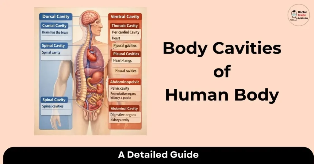
The ventral and dorsal body cavities are the two largest cavities in the human body. The brain and spinal cord are situated in the dorsal body cavity.
What are Body Cavities?
The human body is separated into many bodily cavities, just like the bodies of many other multicellular species. The fluid-filled area inside the body that houses and shields internal organs is called a body cavity.
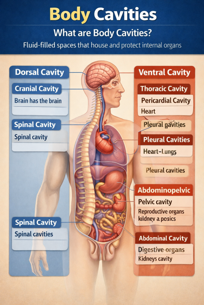
Membranes and other structures divide the cavities in the human body. The dorsal and ventral cavities are the two biggest cavities in the human body. There are smaller bodily cavities inside these two.
Ventral Cavity
The anterior, or front, of the trunk is where the ventral cavity is located. The lungs, heart, stomach, intestines, and reproductive organs are among the organs housed in this bodily cavity. As the organs work, the ventral cavity permits significant changes in their size and shape.
For instance, organs like the uterus, stomach, or lungs can grow or contract without causing other tissues to change or interfering with the functions of neighboring organs.
- The organs within the ventral cavity can undergo significant changes in size and shape as they carry out their activities.
- Organs like the uterus, stomach, or lungs, for instance, are able to expand or contract without causing damage to other tissues or interfering with the functions of neighboring organs.
The thoracic and abdominopelvic cavities are two divisions of the ventral cavity.
- The thoracic cavity, which occupies the chest, is separated into the pericardial cavity and two pleural cavities. The heart is located in the pericardial cavity, whereas the lungs are located in the pleural cavities.
- The lower half of the trunk is occupied by the abdominopelvic cavity, which is separated into the pelvic and abdominal cavities. The kidneys and digestive organs are located in the abdominal cavity, whereas the reproductive and excretory organs are located in the pelvic cavity.
Dorsal Cavity
The posterior, or back, region of the body, which includes the head and the rear of the trunk, is known as the dorsal cavity. The cranial and spinal cavities are two divisions of the dorsal cavity.
- The brain is located in the cranial cavity, which occupies the majority of the upper portion of the skull.
- Inside the vertebral column lies a very long, narrow chamber called the spinal cavity. It includes the spinal cord and extends the whole length of the trunk.
The Meninges
The vertebrae of the spine and the bones of the skull protect the brain and spinal cord. The meninges, a three-layered membrane that surrounds the brain and spinal cord, provide additional protection. Between two of the meningeal layers lies a thin layer of cerebrospinal fluid. The brain produces this transparent fluid, which gives the brain and spinal cord more protection and cushioning.
A bacterial or viral infection is often the cause of inflammation in the meningeal membranes that shield the brain and spinal cord within their cavities. Meningitis is the name for this illness, which, if left untreated, can have major long-term effects, including deafness, epilepsy, or cognitive impairments. Meningitis is considered a medical emergency since it can quickly become life-threatening.
The Abdominopelvic Cavity (Lower Ventral)
The largest area in the body is located under the diaphragm. Due to the lack of a physical boundary dividing the “abdomen” and the “pelvis,” they are frequently combined.
But we separate them functionally:
- The cavity in the abdomen.
- The main digestive organs are located in the “upper” section.
- Digestion in the stomach and intestines.
- Bile storage and metabolism in the liver and gallbladder.
- The immune system’s spleen.
- Kidneys (filtering, although they really occupy a “retroperitoneal” location, somewhat beyond the main cavity lining).
The Pelvic Cavity
The “lower” portion, cradled by the hip bones:
- Urinary Bladder
- Reproductive Organs (Uterus, ovaries, or prostate)
- Rectum
The “Secret” Small Cavities
Although the dorsal and ventral cavities are the “big rooms,” the body also has many smaller, more specialized cavities that are sometimes overlooked:

- The mouth, which houses the tongue and teeth, is called the oral cavity.
- The nasal cavity is a component of the respiratory system that is found inside the nose.
- The “sockets” in the skull where the eyes are located are called orbital cavities.
- The tiny chambers called middle ear cavities are home to the ossicles, which are tiny bones that carry sound.
- Joints (such as the knee) include synovial cavities. They have synovial fluid, which prevents bone-on-bone grinding by acting as a lubricant.
The Protective Lining: Serous Membranes
The friction from breathing or pounding would ultimately rip the tissue apart if your heart and lungs were simply sitting in these dry chambers.
The ventral cavities are protected by serous membranes to stop this.
- These are “bags” with two layers that contain a thin layer of serous fluid:
- The outer layer that borders the cavity wall is called the parietal layer.
- The inner layer that “shrink-wraps” the organ itself is called the visceral layer.
Important Serous Membranes to Understand:
- The pleura envelops the lungs.
- The pericardium envelops the heart.
- Numerous organs in the abdominopelvic cavity are surrounded by the peritoneum.
Why Does This Matter?
Understanding these voids is crucial from a clinical standpoint. When a patient has “peritonitis,” a physician is aware that the abdominal cavity’s lining is inflamed. When someone develops a “pleural effusion,” extra fluid is trapped around their lungs.
Our biology makes sure that an illness in the lungs (thoracic) does not always instantly travel to the brain (cranial) or the stomach (abdominal) by dividing the body into various compartments. It is a biological containment system.