Different level of radiation is measured with different levels of measuring devices or meters in different ways. These radiation dose units help a radiographer or technologist to measure the amount of radiation used, in those particular exposures, with the proper level of radiation dose, with concern for radiation protection to the patient and staff, or the public.
In this chapter, we are going to learn about different types of radiation dose units and their measurements.
What is Ionizing Radiation?
Ionizing radiation is a type of radiation that has enough energy to knock out an electron from any atom or molecule. The loss or gain of electrons is called an ionization, and that charged atom is known as an ion.
Properties: Consider When Ionizing Radiation is Measured
When a radiation dose is measured, many factors are considered to perform the task smoothly, so here are a few properties that are taken into account while measuring the radon exposure:
- Strength of radioactivity of the radiation source,
- Energy and strength of radiation,
- An absorption rate of the targeted materials,
Here, the radiation dose risk is accounted for based on the risk vs benefits factor of the radiation while exposed.
These are taken into consideration while radiation exposure is measured and evaluated.
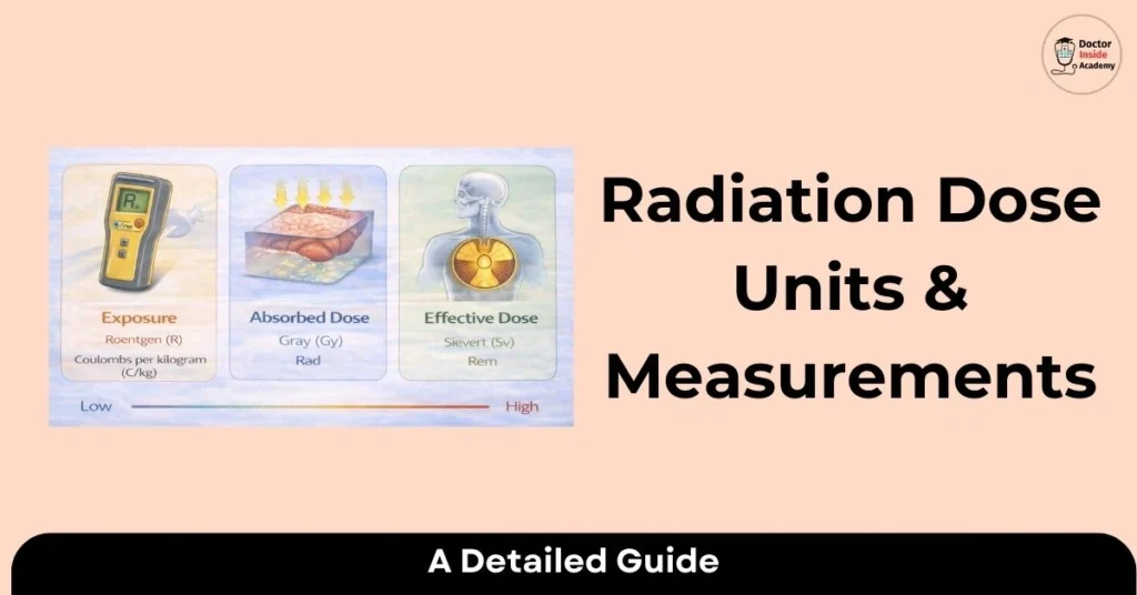
How Radiation Exposure is Measured?
- The human body is exposed to radiation all day, whether from the sun or any man-made source. However, not all reactions are harmful to biological tissues.
- These radiations may be ionizing or non-ionizing. If ionizing, then safety measures must be taken.
- Radiation is absorbed by these tissues and is calculated by a measuring scale named the radiation absorbed dose.
- After that, a specific radiation exposure measurement is known as a radiation-effective dose.
- These X-rays may be X-rays, gamma rays, alpha or beta rays, and are dangerous to biological tissues or organs.
- To measure radiation exposure, there are many units used, such as Roentgen, Sieverts, and Gray.
A Few Common Factors Affecting Radiation Exposure
Radiation is affected by many factors, a few of which are the following:
- The intensity of the radiation,
- How much time irequireden for an exposure?
- Which type of tissue is going to be exposed?
These factors give the idea of the radiation exposure level, and then radiation safety measures are implemented perfectly.
Measuring Biological Risk
Radiation is very harmful to biological tissues or organs; to measure the biological risk of an individual, many radiation measuring doses are used, like rem or Sv.
This measuring factor is based on which type of tissue is exposed at which exposure level.
Measuring Radiation Dose Units and Doses
Many radiation dose measuring units and parameters are used and are given at standard levels or by international levels.
Here are a few of them:
Roentgen
- Roentgen is named after the father of X-rays, Wilhelm Conrad Roentgen.
- Roentgen is the international exposure unit and is used to measure the exposure dose for x-rays or gamma rays.
Gray(Gy)
- Gray is named after British physician L. Harold Gray (1905-1965).
- Gray is a unit of the absorbed radiation dose.
- It is equal to a dose of one joule of energy absorbed per kilogram of matter.
Milligray
- Milligray is a unit of absorbed dose, equal to one thousandth of a gray, or 0.1 rad.
- It is denoted as mGy.
Roentgen Equivalent Man (rem)
- Roentgen Equivalent Man is a unit of radiation measurement to evaluate different responses of biological tissues.
- It is also known as equivalent dose and is denoted by rem.
Millirem
- It is the one-thousandth of rem and is a measuring unit of equivalent dose.
Sievert
- It is denoted by Sv and is known as a radiation effect.
- Sieverts is the unit of ionizing radiation doses, where relative sensitivities of different tissues and organs are exposed.
Miievert
- Milliosievert is one thousandth of a sievert, used for measuring the radiation effective dose.
Becquerels
- Becquerels is the international standard unit of radioactivity. It represents the activity of how much quality of radioactive substance is used at the nucleus that decays per second.
Curie
- Curie isunit of measurement for radioactivity. This unit is named after Marie and Pierre Curie, who studied radium.
- It is defined as 3.7 into 10`10 decays per second and is denoted by Ci.
Picocuries
- Picocurie is one trillionth of a curie.
- The higher the picocurie, the higher the released radiation.
Radiation Dose Quantities
Mostly, these are the main radiation dose quantities used in radiology, which are explained below in an easy-to-understand concept.
Radioactivity
- Radioactivity is the measurement of ionizing radiation released by any radioactive material and the result of nuclear decay.
- Radioactivity is seen as gamma decay, beta decay, or alpha decay.
- Henry Becquerel discovered radioactivity.
Air Kerma
- Air kerma is the simplest unit of radiation and measures the radiation in the air.
- Air kerma is measured by the unit called Gray.
Radiation Absorbed Dose
- Absorbed dose is the amount of radiation absorbed by the tissue, which is proportional to the amount of radiation exposure during the exposure.
- It is also known as RAD and measured as energy deposited per unit mass of the material.
- Absorbed dose is also measured in the Gray unit.
Equivalent Dose
- The equivalent dose is the amount of radiation that may damage the target tissue or organs.
- It is calculated for each organ, with effectiveness, and the type of radiation.
- Different body parts have different levels of radiation absorption and have different densities of the organs or tissues.
- The equivalent dose is calculated in millisieverts(mSv).
Effective Dose
- The effective dose is radiation, where the whole body exposure level is accounted for, with risk vs benefit factors.
- How much radiation dose is effective in getting the best diagnostic result?
- Effective dose is also calculated in millisieverts(mSv).
Difference between Absorbed and Equivalent Dose
The main differences between absorbed and equivalent doses are:
- Absorbed dose is the energy deposited in the small volume of the equivalent organs.
- Where the equivalent dose shows the impact of radiation on these tissues, the type of radiation used is considered.
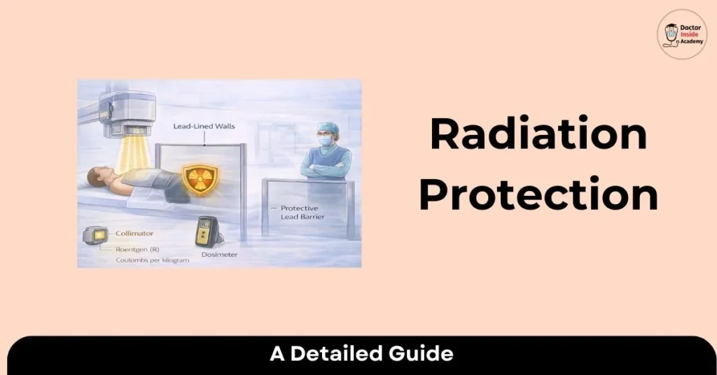
What are the Main Ways to Control Radiation Exposure?
Radiation is very harmful to biological tissue, but after taking a few radiation safety measures, public health was protected.
Here are a few ways that are used to control radiation exposure:
- Proper radiation safety training and education,
- By reducing radiation exposure time,
- By increasing the distance from the radiation source,
- Using proper shielding,
- Proper monitoring of radiation exposure.
Radiation dose is not like a normal absorbed dose of ed, equivalent, or effective dose. So, proper safety measures should be followed before processing any exam or study.
Importance of Informing Patients About Radiation Doses
- Radiation is most commonly used for diagnostic and screening purposes, to evaluate different levels of diseases or abnormalities.
- As this radiation is harmful to tissues, the benefits over risk are followed by the ALARA principle to get the best result.
- The radiology department must discuss with the patient about radiation dose, how this helps them, and the consequences if any contrast study or high radiation dose study is undertaken.
- Public awareness is very important and has been growing in the past dthe to seminars, webinars, and the availability of the Internet for the public.
- Proper patient consent is compulsory before performing any exam in radiology.
- Safety measures are very important and must be followed as per rules and regulations.
Last Words
Now, we can understand different radiation dose units and their measurements, and with this, we discussed a few radiologic doses, most frequently used in the radiology department. With a discussion of patient safety concerns.
We hope you learned something new today. Stay connected for the next chapter.