
As we know, 99 % of the heat and 1% of the radiation are produced when any exposure is taken for diagnostic or screening purposes. Here, nearly 90% of the radiation is not useful, so the remaining one must be utilized to the maximum to get the optimum result. To execute this, intensifying screens are introduced.
So, in this chapter of X-Ray Course Notes, we are going to learn about intensifying screens, how intensifying screens are made, types of Intensifying Screens, layers of intensifying screens, and much more.
Definition of Intensifying Screen
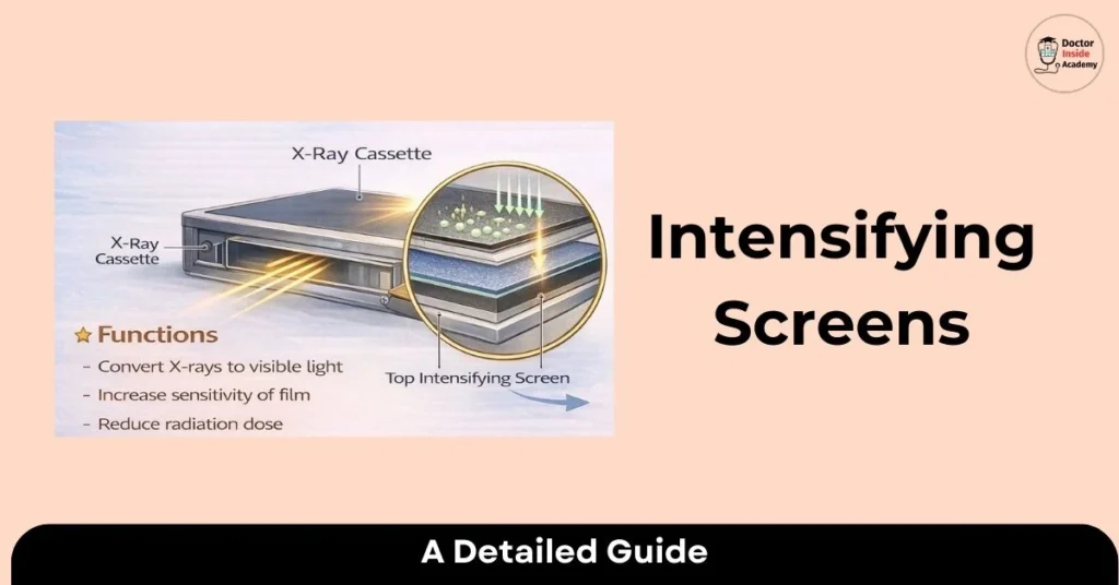
- An intensifying screen is a thin sheet that absorbs X-ray radiation (photons) and converts them into visible light. It is a part of the radiographic cassette and acts as an amplifier for radiation.
- Intensifying screens directly reduce the mAs during the exposure, reducing the radiation exposure for the patients and reducing radiation risks.
- These are mounted on both sides of X-ray cassettes in general radiography, while a single side is used in mammographic exams, mostly on the black side of the X-ray cassette.
Define Intensifying Screen in Dentistry?
- Dental radiographs are used for many diagnostic and treatment purposes for oral diseases, any abnormalities of teeth, or any oral pathology. The radiation used here is very low, and the ALARA principle is strictly followed.
- Along with these, intensifying screens are also used in dental radiography to minimize the radiation exposure to the patients. These intensifying screens in dentistry contain phosphor crystals for the fluorescence effect when radiation is exposed.
Purpose of Intensifying Screens in Radiography
Intensifying screens are used to intensify the effects of the radiation, here; x ray photons.
Let’s discuss a few purposes of intensifying screens as follows:
- to provide a large number of light photons,
- to reduce the exposure duration,
These are the two main purposes of intensifying screens in radiography.
Advantages of Intensifying Screen
In order to lower the patient dosage and improve the quality of the final X-ray image, intensifying screens are quite helpful. These two are insufficient; therefore, let’s talk about a few other benefits of intensifying screens:
Lower Patient Radiation Dose
- The main advantage of an intensifying screen is that it reduces the patient’s radiation dose to a greater extent.
- So, this saves the patient from unwanted radiation exposure and reduces the rate of radiation-related side effects or abnormalities.
Improves Image Quality
- The second most important advantage of intensifying screens is to improve image quality. Along with this, it increases the film sensitivity by improving image contrast and details of the radiographic images.
- These enhanced X-ray images are easy to read and also help radiologists in better diagnosis.
Reduces Radiation Exposure Time
- The third main advantage of intensifying screens is shorter radiation exposure time. The amplification effect of X-ray photons on the X-ray films boosts the X-ray image formation by reducing the radiation exposure time.
- This decreases radiation exposure time and again saves the patient from radiation exposure.
Cost Effective
- Cost-effectiveness is the final but not least benefit of intensifying screens. It lowers X-ray film consumption and maintenance requirements with the aid of intensifying screens. These extend the life of all X-ray equipment and lower maintenance expenses.
- Here are a few benefits of magnifying screens: lowering radiation exposure, enhancing picture quality, and increasing the efficiency and precision of radiological imaging for the radiology department.
Disadvantages of Intensifying Screen
Every coin has two faces; intensifying screens also have their advantages and disadvantages. Here, we already studied a few advantages of intensifying screens, and now we are going to learn the disadvantages of intensifying screens:
Loss of Image Resolution
- The overall X-ray image detail and image resolution are reduced because of the diffusion of light.
- This also blurs the X-ray images, with a reduction in X-ray image sharpness.
Screen Ageing
- Due to regular use of intensifying screens, the overall quality of this screen degrades or tears due to repeated use in repetition.
- This intensifying screen aging leads to a weakening of its overall efficiency, resulting in improper quality of the X-ray images.
Degradation of Phosphor Crystals
- As we know, most intensifying screens contain phosphor crystals. The regular use of these crystals makes them weaker and faded over time.
- This leads to improper sensitivity and light efficiency, with decreased final X-ray image quality.
Effects of Radiation Scattering
- Sometimes, these screens cause radiation scattering, and this leads to degradation of image contrast and also increases the background image noise.
- To decrease the effect of radiation here, we can use collimation or radiation shielding techniques.
So, these are a few disadvantages of intensifying screens, including degradation of the material, loss of image quality, etc. It’s very important to understand all these limitations.
Composition and Materials Used in Intensifying Screens
X-ray film speed is increased while spatial resolution is decreased. An intensifying screen’s 0.4 mm thickness has an impact on both the screen’s speed and spatial resolution.
So, here is a breakdown of all layers of intensifying screens as follows:
Layers of Intensifying Screen
- An intensifying screen contains many layers, designed to convert X-rays into visible light. So, these are the components in a layer of an intensifying screen:
Base layer
- The first layer of an intensifying screen is the X-ray film. Its base layer is made of polyester and is 1 mm thick.
- This layer provides mechanical support to the active phosphor layer. Its base layer must be moisture-resistant, unaffected by radiation, and chemically inert and non-reactive.
Phosphor Layer
- Also known as a luminescent layer. It is the active layer of an intensifying screen.
- It absorbs X-ray photons and converts them into visible light, and this layer contains a substance called ” phosphors”.
- Mostly, the thickness of this is about 50 to 300 μm, and the crystal thickness is about 5 to 15 μm. This layer is crystalline and suspended in polymers, making it flexible.
- The phosphor layer is mostly made up of rare earth metals like Gadolinium, Lanthanum, and Yttrium, and previously, Calcium Tungstate (CaWO4).
A few other types of material used in the phosphor layer are zinc sulfide and barium lead sulfate.
Reflecting Layer
- The reflective layer is between the phosphor and the base layer. The thickness of this layer is about 25 μm.
- It is made up of magnesium or titanium oxide. The reflective layer is made up of TiO2 and is 1 mm thick.
- This layer redirects the radiation; if this is not available, then most of the radiation directly reaches the X-ray film.
A Protective Layer
- The protective layer is composed of a cellulose compound of 0.7-0.8 mm. It is a very close layer to the X-ray film also.
- This layer prevents the intensifying screen from being damaged, is transparent to light, and also provides a cleaning surface.
- Many rare earth elements are used in today’s radiology, as these have high absorption and conversion rates.
Here are a few of them:
- Gadolinium: Green Light
- Lanthanum: Blue Light
- Yttrium
The X-ray film will not absorb or process light if its wavelength is outside of the native range. In this case, orthochromatic films are sensitive to UV, blue, and green light, while conventional films are only sensitive to UV and blue light.
These are components of a layer of intensifying screen, and this combination optimizes the overall X-ray absorption rates and image quality.
Explanation of How Intensifying Screens Work
By lowering the radiation level, the intensifying screen transforms X-ray radiation into light that travels through radiographic film and produces the latent image. This film is utilized in dentistry for cephalometric radiography and panoramic imaging.
Let’s understand how intensifying screens work:
Absorption of Photons
- When the radiation interacts with phosphor material in the layer of the intensifying screens, the radiation is absorbed by atoms of the phosphor material.
Excitation of Electrons
- Now, the absorbed radiation photons cause atomic movement in the phosphor atoms, where the energy level moves to a higher level, and this process is also known as excitation.
Emission of Visible Light
- Here, light photons are released by the excited electrons. These light photons have their wavelength range in the visible spectrum.
Amplification Effect
- These light photons now interact with silver halide crystals in the X-ray film, and this interaction goes with a chemical reaction and starts forming a latent image.
Formation of Latent Image
- After the amplification effect in intensifying screens, there is a presence of a latent image is seen.
Formation of the Final X-ray Image
- Now, this latent image interacts with the photographic emulsion on the film surface, and this process starts forming the final X-ray image, with attenuation of different body parts to show the different anatomical surfaces or body parts.
This overall process of how intensifying screens work produces the final enhanced X-ray image.
With better quality and reduced radiation dose to the patient, also.
Types of Intensifying Screens
These intensifying screens are classified based on the presence of components like phosphor, etc. So, here are a few types of intensifying screens:
Mostly screen uses rare earth elements like gadolinium, lanthanum, or yttrium.
Lead Screens
- The lead screens are placed on both sides of X-ray film, so these are also called the front and back screens.
- Lead screens are made from lead foils; the front screen also acts as a soft radiation absorber and absorbs the scattering of radiation.
- The lead intensifying screen is used mainly in X-rays and gamma radiation.
- The thickness of lead foils is 0.02 to 1.0 mm, depending on which radiation energy is used. Here, the front screen is about 0.02 to 0.20 mm, and the back screen is usually 0.25mm.
- These lead screens are polished so that they are free from surface defects like scratches and will not be transferred to the final X-ray image after processing.
- Lead screen intensifying screens are best suited for radiation levels of 80 keV to 420 keV, and not for high radiation energy.
- These intensifying screens reduce radiation scattering and provide the best X-ray image quality.
- Also improves the image contrast and image quality, with decreased exposure time
Steel and Copper Screens
- In case of high radiation, lead is not the best suited for these intensifying screens; here, steel or copper screens are considered.
- These are used to get better image results in a higher radiation energy range than lead screens.
Fluorescent Screens
- Fluorescent screens work while exposure is ongoing; after the termination of radiation exposure, no fluorescence is seen.
- These are made of a thin base layer, and that base layer contains crystalline metallic salt, usually made from calcium tungstate, which shows fluorescent characteristics.
- This light sensitivity depends on the radiation density.
Flurometallic Screens
- Flurometallic screens have properties that range between the lead screen and fluorescent screen and are made up of a lead-coated foil with a fluorescent layer.
- These have shorter life spans than lead-intensifying screens.
- These also provide appropriate image contrast in the radiographic image while considering reduced exposure time.
- Here, the intensification depends upon the sensitivity of the X-ray film.
Last Words
So, today, we learned all about intensifying screens, how they work, and different types of intensifying screens with their advantages and disadvantages. We hope you enjoyed this chapter of our ” X-Ray Course” by “Doctor Inside Academy”. If there is any doubt or query, feel free to contact our team in the Contact Us section here.