Density is the mass per volume factor that defines how much space any substance occupies with its length, breadth, and height. Simply means the density of any substance is how tightly the particles of that substance are combined.
These densities play a very important role in radiology and are known as radiographic density. As a radiology student, it’s very important to understand these densities in detail. So, in this chapter, we are going to learn about radiographic density and the five types of radiographic densities with their definition, appearance, and examples.
Let’s start learning:
Radiographic Densities
- The radiographic density is defined as the level of darkness in an X-ray image, calculated by the radiation absorption by those tissues in that radiographic image.
- A radiography image appears in different shades of gray, like white, black, greyish, or patchy, or bright white somewhere. These variations are accounted for in radiographic density.
- Radiographic density is also known as radiodensity or the differences in degrees of blackness on an image.
- Along with these, the amount of radiation from the exposure technique is also considered in X-ray image production.
- If exposure is set higher than required, the image will result in overexposure, and if adjusted lower than appropriate, resulting in underexposure of the X-ray image.
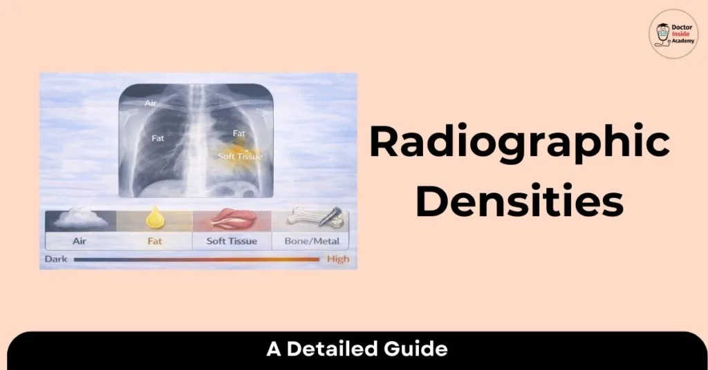
- There are 5 basic radiographic densities in radiology: air, fat, water (soft tissue), bone, and metal. Air is the most radiolucent (blackest), and metal is the most radiopaque (whitest).
Importance of Radiographic Densities in Medical Imaging
- The Radiographic density is very important in medical imaging to visualize the different types of organs and tissues in the X-ray image.
- These are based on different levels of absorption of X-rays by those tissues or organs. This absorption level is based on the composition and density of that organ.
- These 5 radiographic densities are divided into an order of air, fat, soft tissues, bone, and metals.
- Of these 5, four are natural and present in the human body, and the remaining one is metal, which is not present in our body and can be implanted, ingested, or swallowed.
- Along with these, the type of body organ that is going to be exposed and the level of radiation exposure must be calculated to get the best output.
- If the organ is denser, the higher the X-ray is attenuated by that tissue.
- For example, air is less dense, very little radiation is attenuated, but in the case of bone, more radiation energy is attenuated.
- Hence, the final X-ray image depends on the density of the organs or tissue with respect to their structure and thickness or composition of that area, which is known as the contrast of the X-ray image.
- So, the density of the human body depends on which type of organs or tissues are going to be exposed to their composition or structure.
- Based on these, 5 radiographic densities exist. These are explained in detail now.
Five Radiographic Densities
These are five radiographic densities with their definition, appearance, and examples.
Air Density
Definition
- Air is the lowest radiographic density because it absorbs very less x rays.
- Air is also known as a radiolucent material.
Appearance on Radiographic Images
- Air density appears black in a radiography image.
- These appear black because of the minimal absorption of radiation by these tissues.
Examples of Air Density
- An example of air density is air-filled structures, like the lungs or the GI tract.
Fat Density
Definition
- The density of fat is the combination of mass and volume of fat. In radiography, this material absorbs a little bit more radiation than air.
Appearance on Radiographic Images
- The density of fat in an X-ray appears darker than air density and seems whitish-greyish in the X-ray image.
Examples of Fat Density
- Examples of fat density are soft tissues that contain fat portions or muscles, like the liver, and adipose tissues.
Soft Tissue Density
Definition
- Soft tissue has a high aqueous content density. These contain water or fluid.
Appearance on Radiographic Images
- Soft tissue densities appear lighter and medium grey in a radiographic image.
- These have higher density levels as these tissues absorb more radiation than water and air, and along with this, are less dense than lower densities.
Examples of Soft Tissue Density
- Examples of soft density in an X-ray image are muscles, the heart, fluid-filled cavities, abdominal regions, etc.
Bone Density
Definition
- Bones are the highest-density material that is seen in your body.
- These are made up of minerals like calcium, as these minerals absorb more radiation than soft tissues or air.
Appearance on Radiographic Images
- The bone density on X-ray appears white, as these absorb higher amounts of radiation.
- Bone has a higher density because their particles are very close and are of a higher atomic number.
- This is known as radiopaque density or the highest density material.
Examples of Bone Density
- Examples of bone density on X-ray are all bones or cartilage, any vascular calcification or nodules, tumors, or cysts in our body.
Metal Density
Definition
- Metal is not a naturally occurring substance in our body,; it is radiopaque among all materials and appears bright white on an X-ray image. This bright white presentation in an X-ray image is known as metal density.
Appearance on Radiographic Images
- The metallic density on the X-ray image appears bright white.
- These are easily diagnosed as they are very different from adjacent tissues, as they absorb higher levels of radiation than bones.
- Hence, materials with the highest density are also known as radioopaque.
Examples of Metal Density
- An example of metal density in an xray is any metal implants, dental implants, anything hard in pockets or wallets, or ingestion of metal objects like coins, nails, iron stripes, IV contrasts, pacemakers, etc.
- Sometimes, babies swallow coins, which are seen in extra bright white in an X-ray image, and are easily visible and need further assessment to remove them from the body.
- These are the five radiographic densities with their definitions, and appearances in X-ray images with their examples.
- As these create the overall contrast of X-ray images with details, helps a technician or radiologist to study and diagnose these images and take further action to save the life of that patient.
Importance of Contrast in Medical Imaging
- Different types of tissues or organs are seen in the X-ray images based on their different levels of density and composition.
- But sometimes, it’s very difficult to visualize the same structure, like arteries or veins. Here to visualize these arteries andveinss clearly, a radioopaque dye is used.
- These contrast dyes alter the x-ray radiation, and the results are based onther time delay of these floes. So, proper time acquisition is very important to track the best results and to view arteries, veins, or any abnormalities.
- Different types of contracted media used in radiology are gas, liquid, suspension, or powder.
- There are many contrast injection routes in our body, such as the mouth, rectum, or Intravenously and these contrasts are mostly water-soluble.
- Hence, contrast dye is used to enhance the final X-ray image and to visualize the internal organs clearly and smoothly, which leads to the best diagnostic and better treatments.
Last Words
These are the five basic radiographic densities in an X-ray image and help the technologists to interpret these images easily by understanding the level of densities. Different types of diseases or abnormalities are diagnosed based on the radiographic anatomy vs pathological anatomy.