
After taking admission to any course, we need to study its subject matter, do projects, and do internships in that field, and after 3 to 4 years, we will be awarded the desired degree by our recognized university.
Here, I passed out from medical imaging technology and faced many problems in my paramedical career. So, I started Doctor Inside Academy, which is a solution for paramedical students and makes every subject easy to understand in a friendly way.
What is Radiation?
Radiation is energy that flows in waves and particles and has electric and magnetic fields simultaneously, hence called electromagnetic waves. It is present everywhere in our surroundings, coming from both man-made technologies like wireless networks and medical imaging as well as natural sources like the sun and soil.
Radiation plays a very important role in the radiology field and helps healthcare professionals to obtain diagnostic images, where physicians can detect, diagnose, and treat various medical abnormalities and conditions.
In our daily lives, radiation – whether from natural or artificial sources – offers numerous benefits. Any body or item that is exposed to radiation can absorb it and scatter it. The majority of natural radiation cannot be harmful to a person’s health, and the sun is one source of this radiation. By removing electrons from atoms’ shells, radiation can ionize them, which can damage biological tissues.
Radiation is classified as either ionizing (high energy, like X-rays) or non-ionizing (low energy, like radio waves) based on its energy level. Radiation as a whole is an important tool in current health, worldwide communication, and the generation of clean energy; even high-energy radiation requires careful safety precautions in order to protect living tissue.
Types of Radiation
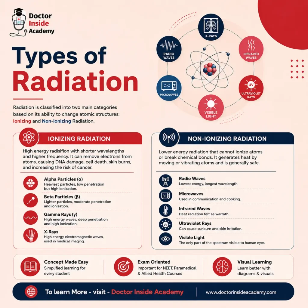
Based on its capacity to change atomic structures, radiation is divided into two main categories: ionizing and non-ionizing radiation. Non-ionizing radiation often merely moves molecules or produces heat, while ionizing radiation has enough energy to break atomic bonds and perhaps damage living tissue.
Ionizing Radiation
Ionizing radiation has high energy because of its shorter wavelengths and higher frequency.
Because of its huge energy, it may drain stable atoms of their electrons, converting them into charged particles known as ions. This can cause serious skin burns, damage DNA, kill biological cells, and increase the long-term risk of cancer if it takes place within the human body.
Ionizing radiation includes the following, based on their interaction levels:
- Alpha Particles
- Beta Particles
- Gamma Rays
- X-Rays
Non-ionizing Radiation
The lower-energy portion of the spectrum is occupied by non-ionizing radiation. It doesn’t have the energy required to break chemical bonds or ionize atoms. Instead, it generates heat by safely shifting or vibrating atoms.
Even though they are usually harmless, prolonged or severe exposure to some varieties might nevertheless result in mild tissue irritation or skin burns.
This non-ionizing radiation is also divided into further categories, are followings, based on their energy levels are;
- Radio Waves
- Microwaves
- Infrared Waves
- Ultraviolet Rays
- Visible Light
What are X-rays?
X-rays are a high-energy form of electromagnetic radiation used extensively in medicine to look inside the human body. They exist in the ionizing part of the electromagnetic spectrum, which means they have enough energy to pass through solid matter as well as to have electric and magnetic characteristics.
Wilhelm Conrad Röntgen (commonly written Roentgen), a German scientist, made the accidental discovery of X-rays in 1895. He noticed a nearby chemically treated screen glowing while experimenting with electrical currents in vacuum tubes, even though the tube had been covered in thick black cardboard. He called these very penetrating beams “X-rays” (using the mathematical symbol “X” for an unknown variable) since the nature of these rays was entirely unknown at the time.
X-rays are produced by passing an electric current through a vacuum tube containing two electrodes: the anode and the cathode.
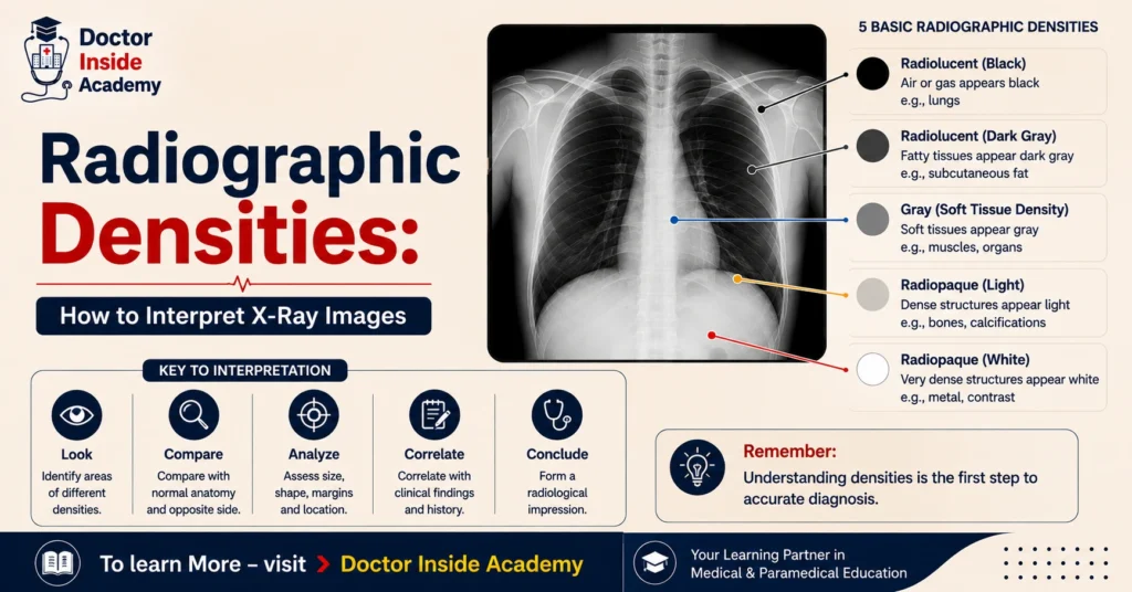
When these X-rays are passed through the human body, they may be absorbed, scattered, or reflected by the medium, depending on their densities and interactions.
Calcium blocks X-rays and appears white on the X-ray images, and the black region that appears on the images shows air, and a few gray shades show fluid, blood, or soft tissues.
What are X-rays used for?
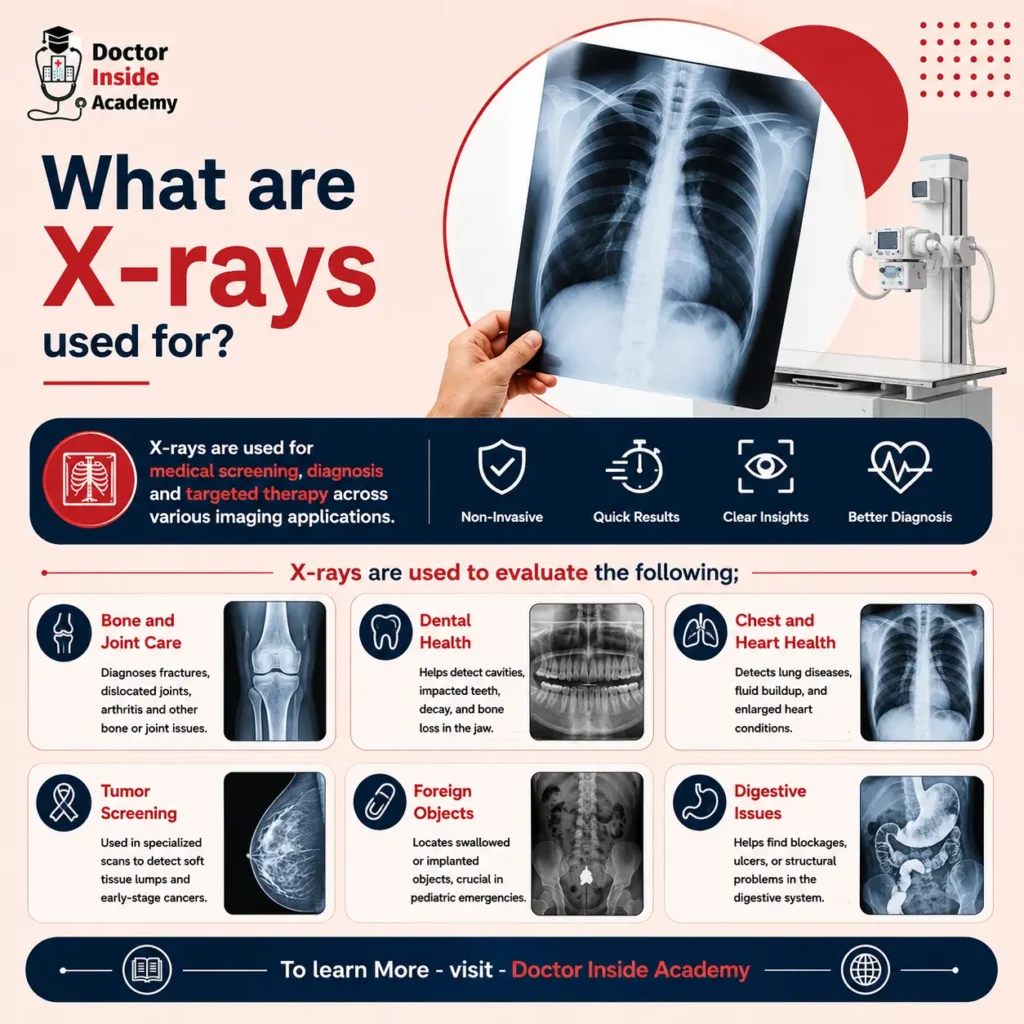
A key component of modern healthcare is X-rays, which are commonly used for medical screening, diagnosis, and targeted therapy across a variety of imaging modalities. They give medical professionals quick and vital insights into a patient’s condition by enabling non-invasive visualization of interior structures.
X-rays are used to evaluate the following;
- Bone and Joint Care: Quickly diagnose fractures, dislocated joints, fractured bones, and arthritis-related damage to joints.
- Dental Health: Used by dentists to identify cavities, impacted wisdom teeth, hidden dental decay, and loss of bone in the jawbone.
- Chest and Heart Health: Identifies fluid buildup, an enlarged heart (cardiomegaly), and lung diseases (such as pneumonia or TB).
- Tumor Screening: Used in specialized scans to find soft tissue lumps, such as early detection of breast cancers.
- Foreign Objects: This function, which is particularly important in pediatric emergency treatment, quickly locates any ingested or implanted objects inside the body.
- Digestive Issues: Assists in locating obstructions, ulcers, or structural problems in the digestive system
X-rays are detected by X-ray cassettes, then converted into hard copies through printers and software. X-rays are used in different modalities like X-ray machines, CT, fluoroscopy, mammography, etc. X-rays are avoided during pregnancy because this will lead to birth defects or other severe abnormalities in the offspring.
Properties of X-Rays
The distinctive physical, electrical, and mechanical characteristics of X-rays control their behavior in a vacuum and their interactions with matter. These special qualities make them ideal for deep-penetration imaging, but they also necessitate stringent safety precautions.
The 12 Key Properties of X-rays are as follows:
- Electromagnetic Nature: They are a high-energy form of electromagnetic radiation.
- Invisible: They completely bypass human vision and remain invisible.
- High Penetration: They possess massive penetrating power through opaque matter.
- Spectrum Profile: They feature short wavelengths (\(0.01\) to \(10\text{ nm}\)) and high frequencies.
- Constant Velocity: They travel at the exact speed of light in a vacuum (\(3 \times 10^8\text{ m/s}\)).
- Massless Particles: They are pure packets of energy and carry absolutely no mass.
- Linear Propagation: They always travel forward in a perfectly straight line.
- Field Immunity: They carry no charge, meaning they cannot be affected by magnetic or electric fields.
- Ionization Ability: They pack enough energy to forcefully ionize atoms and molecules.
- Biological Risk: Because of ionization, they are destructive and harmful to biological tissues.
- Imaging Capability: They chemically interact to produce clear images on photographic films.
- High Voltage Creation: They require a high-voltage current to form inside an X-ray tube.
Different Types of X-ray Studies
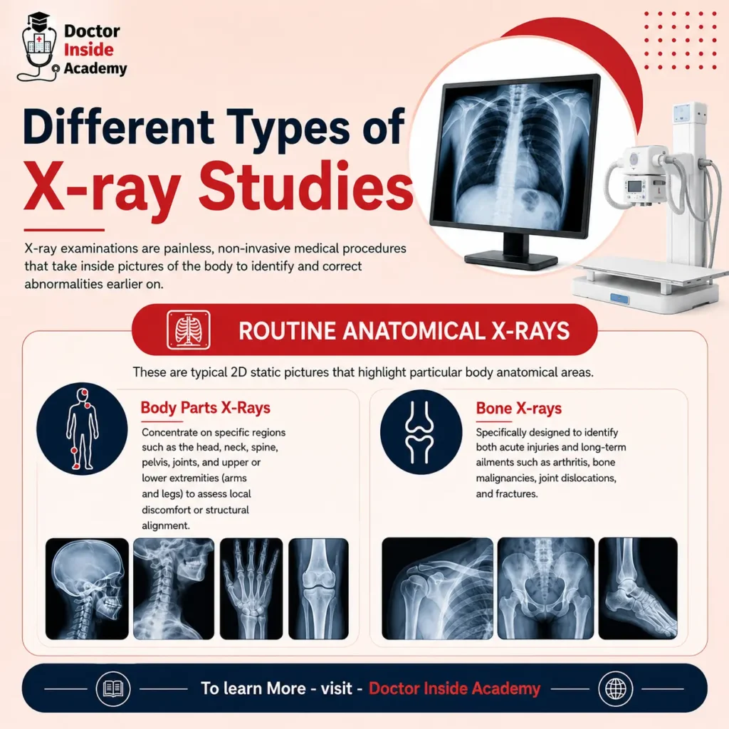
X-ray examinations are painless, non-invasive medical procedures that take inside pictures of the body to identify and correct abnormalities earlier on. Medical professionals can examine anything from solid bone structures to moving interior organs using a variety of methods and tools.
Routine Anatomical X-Rays
These are typical 2D static pictures that highlight particular body anatomical areas:
- Body Parts X-Rays: In order to assess local discomfort or structural alignment, body parts X-rays concentrate on specific regions such as the head, neck, spine, pelvis, joints, and upper or lower extremities (arms and legs).
- Bone X-rays: Specifically designed to identify both acute injuries and long-term ailments such as arthritis, bone malignancies, joint dislocations, and fractures.
- Chest X-Rays: One of the most popular medical scans is a chest X-ray, which shows the heart, lungs, ribs, and diaphragm. It is used to identify lung cancer, pneumonia, TB, rib fractures, and chronic coughs.
Abdominal and Specialized Urinary Scans
These studies assess the soft tissue architecture of the digestive and urinary systems by looking beyond the skeletal system:
- Abdomen X-Rays: X-rays of the abdomen show the stomach, intestines, liver, spleen, kidneys, and bladder. It is often used in conjunction with contrast investigations (such as a barium enema) to highlight the digestive tract and aids in the identification of intestinal obstructions and internal organ damage.
- KUB X-Rays (Kidneys, Ureters, and Bladder): An examination of the abdomen that focuses on the urinary system. It is mostly used to find kidney or ureteral stones and assesses the size, shape, and location of these organs.
Dedicated Screening and Target-Specific X-Rays
These techniques assess extremely specific tissues using advanced technology and positioning:
- Dental X-rays: Take images of the teeth, gums, and jawbone to find cavities and detect illnesses. The OPG (OOrthopantomogram, which offers a panoramic, ear-to-ear view of the whole mouth, is the most popular kind.
- Mammography: A low-dose X-ray machine made specifically for the female breast. To find breast lumps, tissue nodes, and breast cancer before physical symptoms manifest, it is utilized for routine screening or diagnostic follow-ups.
Advanced 3D and Dynamic Modalities
The X-ray beam is modified by modern technologies to record improved dimensions or motion in real time:
- Computed Tomography (CT Scan): Multiple cross-sectional slices are taken by rotating an X-ray source around the body in a computed tomography (CT) scan. It provides significantly greater visibility than standard 2D radiography pictures by creating intricate 3D views of organs, soft tissues, and blood vessels.
- Fluoroscopy: Creates moving pictures of inside organs in real time, much like an “X-ray movie.” It enables medical professionals to see dynamic body processes, such as the mechanics of eating or the movement of contrast dye via blood vessels.
X-Ray Contrast Studies
Specialized contrast media are used in X-ray contrast studies to highlight particular interior organs, blood vessels, and tissues in high-definition detail. Soft structures that would normally be translucent on a typical X-ray scan become apparent because these contrast agents alter how X-rays interact with the body.
Contrast Media Basics and Administration
Contrast medium improves the visible contrast of interior structures by acting as a temporary dye.
- Physical Forms: Contrast media can be liquids (such as iodinated contrast), suspensions (such as barium sulfate), or powder gases (which are used to inflate organs with air in double-contrast examinations).
- Routes of Administration: The target area determines how the medium is supplied.
- Oral: Swallowed to assess the upper digestive system.
- Rectal: To observe the lower large intestine, an enema is used.
- Intravenous (IV): Directly injected into blood vessels to monitor the urinary and circulatory systems.
- Direct Injection: Directly injected into the urethra, cervix, or joints.
- Risk vs. Benefit: Contrast studies are a risk vs. benefit process, no matter their great efficacy. Iodinated dyes may cause allergic responses, nausea, or renal strain in certain people; thus, doctors must carefully review the patient’s medical history, particularly with regard to kidney function, before moving further.
Gastrointestinal (GI) Contrast Procedures
To identify structural anomalies, these investigations follow a thick barium liquid as it covers the digestive system lining:
- Esophagram (Barium Swallow): The patient drinks a barium solution while a radiologist takes both still and moving fluoroscopy pictures to assess the pharynx and esophagus’s structural integrity and swallowing mechanics.
- Upper GI Series: A longer series of X-rays that follow the contrast throughout the stomach, esophagus, and first segment of the small intestine to identify tissue inflammation, tumors, ulcers, and hiatal hernias.
- Small Bowel Series: A timed X-ray scan that monitors intestinal transit and obstructions by tracking the barium as it passes completely through the small intestine and approaches the big intestine.
- Barium Enema: To coat the colon and rectum, contrast is injected rectally. Diverticulitis, polyps, colon cancer, aberrant tissue passageways (fistulas), artificial colon dilatation, and structural blockages are all diagnosed with this research.
Circulatory, Reproductive, and Joint Procedures
Direct contrast injection is necessary for these extremely specialized techniques to map interior cavities and narrowed pathways:
- Angiography: To map the blood vessels in the heart, lungs, kidneys, brain, arms, or legs, an IV contrast agent is delivered directly into the circulatory system. It is essential for the diagnosis of aneurysms, blood clots, vascular tumors, and artery obstructions.
- Hysterosalpingography (HSG): The reproductive tract is softly injected with contrast material via the cervix. This X-ray scan is a main diagnostic technique for examining infertility or blockages since it assesses the structural health of the uterus and fallopian tubes.
- Arthrogram: To identify hidden cartilage tears, ligament injury, or abnormalities in the joint capsule in the arms or legs, contrast media is injected directly into a joint area (such as the shoulder, knee, or hip).
Urinary Tract Contrast Procedures
These investigations use iodinated contrast dyes to separate the urinary system’s filtration and elimination pathways:
- Intravenous Pyelogram (IVP): Contrast is injected into an arm vein, filtered by the kidneys, and eliminated through the urine in an intravenous pyelogram (IVP). In order to determine kidney stones, cysts, tumors, or an enlarged prostate impeding urine flow, timed X-rays record the dye passing through the kidneys, ureters, and bladder.
- RGU / MCU (Retrograde Urethrogram / Micturating Cystourethrogram): The male urethral area is the focus of very specialized contrast examinations called RGU/MCU (Retrograde Urethrogram/Micturating Cystourethrogram). While MCU takes X-rays while the patient empties their bladder to look for irregular backward urine flow (reflux), RGU flushes contrast backward up the urethra to discover narrow strictures.
Precautions During Use of X-Ray Contrast Media Before and During the Procedure
Certain pre-scan guidelines must be followed strictly to ensure patient safety and avoid image distortions (artifacts):
- Fasting: Before abdominal or pelvic contrast scans, patients are usually told to fast (not eat or drink) for a few hours. This lessens the possibility of contrast-induced nausea and ensures that the stomach and intestines are free of debris.
- Metallic Clearances: All jewelry and metal items must be taken off the body entirely. Metal completely filters X-rays and creates dazzling white shadows that can mask important diagnostic information.
- Bladder Preparation: To provide clear imaging of the urinary structures, patients may be advised to empty their bladders right before the operation begins, depending on the research (such as an IVP or MCU).
- Motion Control: When the technologist gives commands, the patient must stay still and hold their breath. A re-scan is required because minor motions might cause the X-ray image to become blurry.
- Mandatory Medical Reporting: Prior to receiving the dye, patients must immediately inform the radiology staff of any current medical issues.
- Allergies & Asthma: The risk of a pulmonary or systemic hypersensitivity reaction to the contrast is greatly increased if you have a history of severe allergies, asthma, or COPD.
- Pregnancy: Potential or confirmed pregnancy must be notified so that alternate imaging or protective shielding can be employed since ionizing radiation carries a risk of birth abnormalities.
- Diabetic: Very important to disclose because some diabetic drugs might seriously impair metabolic systems when used with contrast media.
Risks and Complications of Contrast Media
While most contrast procedures are safe, there are potential risks when foreign drugs are introduced into the circulatory or digestive systems:
- Severity of Responses: There are three levels of adverse physiological responses to contrast dyes:
- Mild: Small problems include a metallic taste in the mouth, nausea, vomiting, or localized warmth.
- Moderate: More noticeable symptoms such as intense itching, slight wheezing, or widespread hives (urticaria).
- Severe: Life-threatening situations that need quick medical attention, such as severe dyspnea, angioedema (facial swelling), a sharp decrease in blood pressure, or anaphylactic shock.
- Kidney Condition: The renal system filters contrast media out of the body. Contrast-Induced Nephropathy (CIN), a kind of transient or permanent kidney injury, is far more likely to occur in patients who already have renal issues.
- The Diabetic Risk Factor: Patients with diabetes are particularly vulnerable to problems from contrast. The contrast dye may hasten serious renal problems in a diabetic patient with underlying kidney disease. Moreover, combining iodinated contrast with popular diabetic drugs (such as metformin) without making the necessary fasting changes might result in a hazardous accumulation of blood acid (lactic acidosis).
- Gastrointestinal Side Effects: Although barium-based contrast agents used in GI examinations are not absorbed by the body, if the barium solidifies inside the intestine following the treatment, they may induce localized digestive disturbances such as vomiting, cramping in the stomach, and severe constipation.
Applications of X-Rays
Because of their exceptional penetrating power, X-rays are a very adaptable type of high-energy electromagnetic radiation that is used in heavy industry, science, health, and security. Through their ability to flow through opaque materials and interact with matter at the atomic level, they enable experts to safely view interior structures, eliminate malignant cells, and examine the basic components of nature.
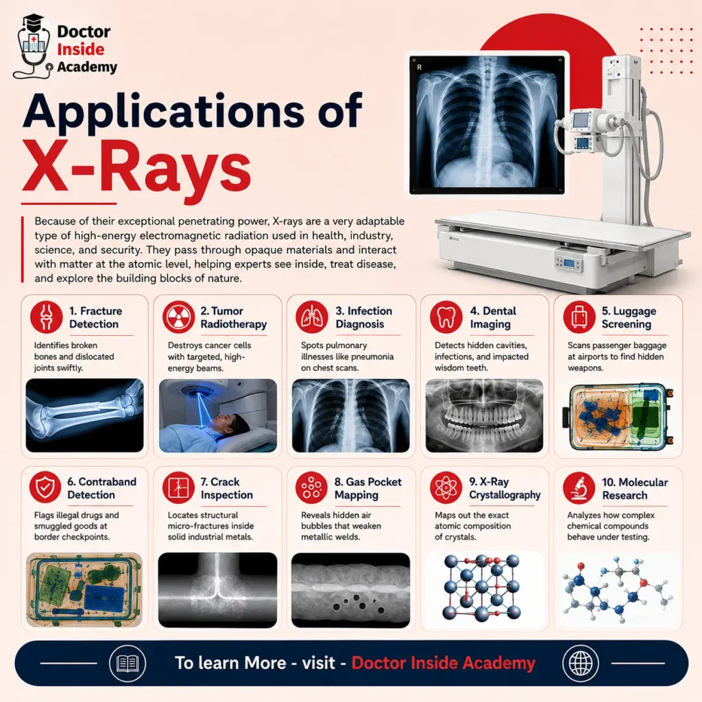
- Fracture Detection: Identifies broken bones and dislocated joints swiftly.
- Tumor Radiotherapy: Destroys cancer cells with targeted, high-energy beams.
- Infection Diagnosis: Spots pulmonary illnesses like pneumonia on chest scans.
- Dental Imaging: Detects hidden cavities, infections, and impacted wisdom teeth.
- Luggage Screening: Scans passenger baggage at airports to find hidden weapons.
- Contraband Detection: Flags illegal drugs and smuggled goods at border checkpoints.
- Crack Inspection: Locates structural micro-fractures inside solid industrial metals.
- Gas Pocket Mapping: Reveals hidden air bubbles that weaken metallic welds.
- X-Ray Crystallography: Maps out the exact atomic composition of crystals.
- Molecular Research: Analyzes how complex chemical compounds behave under testing.
How Does X-Ray Radiation Affect the Human Body?
Ionization, the process by which high-energy photons impact atoms and remove their electrons, is how X-ray radiation affects human health. This disruption of cells begins at the molecular level, when DNA strands are damaged and chemical connections are broken. This damage can change cells, harm tissues, lead to organ failure, and somewhat raise the long-term risk of cancer if the body is unable to heal it.
How X-Rays Affect Specific Body Parts?
- Bone Marrow: High sensitivity; radiation can temporarily reduce blood cell numbers by depleting blood-forming stem cells.
- Reproductive Organs: Extremely sensitive; exposure may interfere with quickly dividing sperm or egg cells and result in temporary sterility.
- Thyroid Gland: Lead thyroid shields are frequently utilized because of the thyroid gland’s high susceptibility to radiation-induced mutations.
- Skin: Moderately sensitive; prolonged or high dosages may result in radiation burns that resemble sunburns, redness, or irritation.
- Eyes: Moderately susceptible; over time, cataracts may develop due to the lens’s susceptibility to cumulative radiation.
- Hair: Reduced sensitivity; localized hair loss may result from severe, highly focused radiation doses (such as those used in cancer treatment).
- Blood Circulatory System: After severe, extensive exposure, white and red blood cell counts may temporarily decline.
- Brain: Reduced sensitivity; mature brain cells are very resistant to typical diagnostic dosages since they do not proliferate quickly.
Cellular and Long-Term Effects
- DNA Damage: X-rays can directly break DNA strands or produce dangerous free radicals that assault genetic material chemically.
- Cancer Risk: Although this danger is extremely unlikely for routine diagnostic scans, irradiated cells that survive with damaged DNA have the potential to mutate and develop cancer later in life.
How were X-rays named?
The mathematical variable “X” was used to denote X-rays in order to indicate that their actual nature and characteristics were entirely unknown. Wilhelm Conrad Röntgen, a German scientist, was unable to quickly identify the kind of radiation when he unintentionally found these extremely penetrating rays in 1895. In his first research report, Röntgen briefly called them “X-rays” since mathematicians typically use the letter “X” to denote an unknown number or variable.
The mysterious and memorable word “X-rays” remained in common English, even though many of his scientific contemporaries later proposed calling them “Röntgen Rays” in his honor, a term that is still used in several languages today.
How Do X-rays Work?
X-rays create an internal image based only on the different densities of your tissues by sending a high-energy electromagnetic radiation beam throughout your body. A heated cathode within a customized vacuum tube releases electrons that are forcefully accelerated by high voltage until they collide with a tungsten anode, where their kinetic energy is transformed into X-ray photons.
While low-density structures like soft tissues, fluids, and air permit the rays to pass through with little to no resistance, solid structures like calcium-rich bones absorb the radiation as it travels throughout a patient. A digital detector or photographic film positioned behind the patient is struck by the residual radiation, producing a grayscale “shadowgraph” that illuminates various body parts according to the amount of radiation that was able to pass through them.
- Electron Generation: A cloud of negatively charged electrons is released by the heated cathode filament.
- Photon Production: When fast electrons crash with a tungsten anode, their kinetic energy is transformed into X-ray photons.
- High-Density Absorption: The rays are absorbed by calcium-rich bones, which keeps them from getting to the film.
- Low-Density Transmission: Air-filled lungs, muscles, and fat are all easily penetrated by radiation.
- White Image Profiles: Dense bone-blocked areas look dazzling white because they are shielded from radiation.
- Black Image Profiles: The detector is completely exposed by rays traveling through empty air, making it completely black.
- Gray Image Profiles: A variety of gray tones are produced by intermediate soft tissues absorbing some radiation.
Conclusion
In this chapter of the X-ray course series, we learned about X-ray radiation and its types, and all about X-rays and their properties, with different types of contrast or non-contrast studies being performed day to day in the radiology department, and radiation protection and safety measures.
I hope you enjoyed this article and are on your way towards your academic career, and you can connect with us on all social media.