
Radiography is another concept of photography, where we use x ray radiation to record images of inner organs or tissues for different diagnostic or screening purposes. For a student, it’s very important to understand the concept of radiographic films or x ray films, to perform best in their academics or practicals.
So, in this chapter, we are going to learn about radiographic films or x ray films, their types, and structure with advantages and limitations.
Radiographic Film
Radiographic film is also known as x ray film and is a very basic component of medical imaging for diagnostic purposes.
X Rays films are used to visualize organs or structures of different body organs or tissues to diagnose diseases or abnormalities.
What is X Ray Film?
X ray films are like photography images because the x ray film represents the content; i.e.,- human body parts or tissues, that are exposed by the x rays.

X ray films are composed of different layers, like a base layer material with base material (polyester), and a protective coating.
What is a latent image?
The latent image is the invisible x ray films, which need to be processed further to make a final x ray image output. These unprocessed images are called latent images.

Types of X Ray Images
X ray images are divided into many types based on their visibility, quality, etc, and are divided into two parts based on the image processing, which are the following:

Latent Image in Radiology
The latent image is invisible and is visible after the film is processed completely.
Latent Image Formation in X Ray
The latent is formed when an x-ray strikes the film receptor after passing through the human body, this film receptor is made up of silver bromide crystals with a polyester base material, these crystals get fluorescent and form the latent image.
Now, the latent image is processed further and developed into x ray image by a step-by-step process called x ray film processing, where this film is developed, rinsed, fixed, washed, and then dried to make the final x ray image.
Final X Ray Image
The final image is a visible x ray image, presented in 2D, with different levels of densities.
The latent image is converted into the final image by a process called image processing.
Structure of X Ray Film
The structure of x ray films contains different layers with their specific functions as x ray films are used to visualize x ray images.
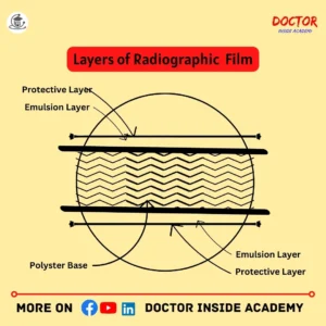
The layers are explained below;
X Ray Film Composition
The x ray film composition contains five layers, which are explained below;
X Ray Film Base
The x ray film base is a central layer of x-ray films, with emulsions on both sides.
The base must be strong enough and flexible, to be used again and again during exposures.
The base layer is made up of cellulose triacetate or polyester and is used for support and provides stability to the x ray film.
Emulsion Layer of X Ray Film
The emulsion layer of x ray film contains gelatin and silver halide.
The gelation here is used to support silver halide crystals and prevent clumping and is very sensitive to radiation and the latent image is formed here.
The emulsion layer also contains a few hardening agents, where the latent image is stored.
As the x-ray film is coated with an emulsion and a base of polyester, the emulsion is on both sides of the base layers with a protective layer on both sides.
Substratum Layer of X Ray Fim
The substratum layer of x ray film is thin and contains gelation and solvents, that are further used to bind the emulsion and the base.
The substratum layer of x ray film is also known as the adhesive layer, as the emulsion and base layer cannot stick to each other, so this is used to keep them binded.
Why do we use Gelatin as a Binder in X-Ray Film?
Gelatin is used as a binder in x-ray film because of the following;
Gelatin has collagen fiber.
Gelatin is a medium where Silver Nitrate and Sodium Bromide react and form AgBr and get dispersed and still suspended.
In a warm state, Gelatin can easily spread and on cooling, it forms a gel on the base layer.
Gelatin is very flexible and cannot react with silver halides.
Protective Layer of X Ray Film
The protective layer of x ray film is the topmost layer and is made up of gelation and protects the emulsion from physical damages like handling, and keeping, and environmental factors also like fogging, different temperatures, etc.
It also prevents x ray images from scratches or chemical degradation.
Anti Halation Layer
The anti halation layer is used to prevent radiation scattering and reduces the risk of image blurring.
The anti halation layer is presented under the emulsion layer.
X Ray Film Speed
The x ray film speed is defined by relative sensitivity to a given amount of radiation and no number or metrics is used to determine the speed of x ray film, not like photographic camera films.
Most films are designed as standard, fast, and ultra-fast.

The total thickness of the radiographic film is about 0.25mm usually and contains different layers, as we already discussed above.
Hence, these layers are designed carefully to ensure the best output with appropriate x-ray absorption. The process of x ray imaging starts by exposing the x ray film, where x rays penetrate the human body and create a latent image in the emulsion layer, where films are developed using a series of film processing, which will discussed later; making that latent image visible x ray image.
So, these are structures of x ray film layers, as x ray films are very important components in medical imaging technology, that are used to capture and visualize images.
Unexposed X Ray Film Storage
The unexposed x ray film must be stored properly and carefully, to make its life longer and for better outputs.

Common Factors, to Keep in Mind While Storing Unexposed Films
- Temperatures must be kept below 24 degrees Celsius.
- Keep unexposed x ray film away from chemicals.
- Stacked them properly,
- The relative humidity of unexposed x ray film must be below 60 degrees Celsius.
The unexposed films must be away from heat and stored in a cool and dry place because the high temperature may damage the x ray films or make them foggy.
If films are kept long in unsuitable areas, fog will develop there; so, sensitive materials must be stored in a cool and kept away from a dry room, and walls must be lined with 105 mm thickness.
The x ray films must be prevented from direct radiation exposure, otherwise, these will be partially exposed and become unable to perform the main exam properly, here, to prevent the x ray films from direct radiation exposure, the lead covering is used or must be kept away from the x-ray room with proper maintenance and care.
If films are kept in hot, tropical, or humid climates, then films are kept in cold storage and if the box is open then films cannot be put back in that cold storage.
X Ray Films be maintained with proper pressure, otherwise, higher pressure leads to bending, cracking, etc will result in image artifacts.
To keep the unexposed x ray film safe from pressure, these will be placed smoothly with proper arrangement.
The emulsion layer of x ray film is very sense too, so film boxes must be kept carefully and keep them safe from radiation exposure with proper maintenance and guidance.
If the large stock of unexposed x ray film is kept for a long time, then it must be kept properly and used according to their expiry date, to make it cost-effective and prevent them from decaying or accidents.
X Ray Film Processing
X ray film processing is a collection of step-by-step procedures to convert latent images into visible x ray images.
To study the latent image, that is invisible to human eyes, many steps are used to develop the final x ray films; so, here are five steps of x ray film processing in radiography., which may be manual or automatic.
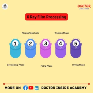
X Ray Film Processing is Done by Five Steps
- Developing,
- Rinsing,
- Fixing,
- Bathing,
- Drying,
- Developing
The latent is converted into a visible x ray image is known as the development of the film and is processed in a solution called developer solution.
The exposed radiographic films are immersed in the chemical developer solution in the developing phase of the x-ray film.
This developing solution results in the latent image in metallic silver and creates a dark area on the x ray film.
Developing Agents
The developing agents used in x ray film processing are Monol, Pyrazon, and Hydroquinone.
Here is other substances in the developer solution are sodium carbonates, sodium hydroxide as an accelerator; sodium bisulfite, sodium sulfite as a preservative; potassium bromide as a restrainer, water softener or gelation harder as additives, used ion a developing solution for better results.
- Rinsing/Stop bath
The next step in x ray film processing, after the film development, is rinsing, here the film is washed and then goes for the next step otherwise it leads to uneven fixation.
The film is washed in a stop bath and this process is also known as rinsing of film, to remove the developer solution.
The rinsing solution in x ray film processing contains approx. 3% solution of acetic acid.
If the rinsing or stop bath, is not done, it will result in x ray images with stain formation on it.
- Fixing
The fixing of the X-ray film is done because it removes silver halides, that are not contributed in an x ray image.
After the developing phase, the x ray film is immersed in the fixer solution, which removes the unposed silver halides crystal and post-image image or permanent x ray image.
The fixing bath contains the solution of sodium thiosulfate, and this solution converts silver halide into a soluble compound.
Here, sometimes ammonium thiosulphate in case of quick fixing.
- Washing
The films will be washed in batches.
Mostly the wash tank contains two chambers, one for films directly coming from fixing and the other for after the fixing bath, and the other one contains fresh water for the smooth wash of film.
- Drying
After the completion of the washing and fixing process, the x ray films are dried, so that chemicals or moisture goes away and helps in better diagnostics of diseases or abnormalities.
Manual drying or air drying is used for this purpose.
Time taken during X Ray Film Processing in Radiography
Here is the average time taken during X-ray film processing in radiography are followings;
- The development process takes approximately four to five and a half minutes; (4 to 5.5 minutes) to convert the latent image into visible image.
- The rinsing process takes about 30 seconds to remove the excess chemicals used during the film’s development.
- The fixation process takes about 10 minutes, to remove the unexposed silver halide from the emulsion.
- The washing process process takes about 10 minutes, and the excess chemical is removed again, after fixation.
- The drying process takes about 30 minutes, during which extra water is removed and x-ray images are used as final radiographs for diagnostic or screening purposes.
Types of Radiographic Film
Radiographic film or x ray films are divided into different types, based on their type and uses in specific clinical applications, are following:
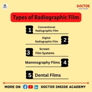
- Conventional Radiographic Film
Conventional radiographic film is used for general radiography, such as chest X-rays, abdomen X-rays, extremities X-rays, etc, where the X-ray images will be processed manually as discussed above in X-ray film processing.
- Digital Radiographic Film
Digital radiographic films can’t require x ray cassettes as these have indirect flat detectors, known as Digital Image Capture Devices; and no manual efforts are done to make these images, as these are directly transferred to the system.
- Screen Film Systems
The screen film system is embedded with intensifying screens and these screens emit light when exposed to radiation, which in return, exposes the x-ray film.
Screen film systems are mostly used in mammography or fluoroscopy.
- Mammography Films
The mammography film is a combination of both; a single emulsion and a single intensifying screen.
Mammography exams are done for diagnostic or screening purposes to study female breast tissues and the most common reason for this is female breast cancer.
- Dental Films
The dental films are mostly wrapped with moisture and light-proof coverings.
The dental x-rays are done to check cavities, tooth alignments, or for planning for braces implantation or oral surgeries.
Conventional Radiographic Film
Conventional radiographic films are also called traditional radiographic films and are mostly used for general radiography, like capturing the chest, abdomen, and lower or upper extremities, mainly.
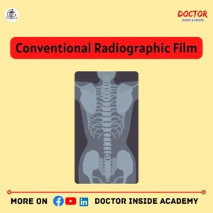
The x ray images are produced manually in conventional radiographic films, mostly steps followed like development and film processing.
Types of Conventional Radiography
Conventional radiography is also further divided into many types based on the type of study, to visualize and diagnose different body parts, are following:
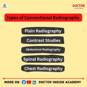
- Plain Radiography
Plain radiography is the most common type of conventional radiography.
Plain X-rays are used to see inside bony structures, find fractures, or evaluate lung conditions like pleural effusion, pneumonia, pneumothorax, etc.
- Fluoroscopy
Fluoroscopy is a concept of taking continuous X-ray images to see the internal organs in real-time during the procedure.
The fluoroscopy is performed when we need to see the live motion of organs, like the heart, throat, and digestive systems followed by barium studies.
- Contrast Studies
The contrast study is a method of taking x ray images, with a dye name contrast, to see the internal organs clearly and highlight them differently than the adjacent organs. For example – IVP, RGU, MCU, and Barium studies like barium swallow, barium follow-through, or barium enema.
- Abdominal Radiography
Abdominal radiography is done to see the organs of the abdomen or tummy regions to evaluate or diagnose the intestines and stomach with their adjacent organs and tissues.
- Spinal Radiography
Spinal radiography includes the X-ray images of all vertebrae, divided into cervical X-rays, thoracic X-rays, lumbar X-rays, or sacrum or coccyx X-rays.
This is done to diagnose or check injuries, slipped discs, back pains, etc.
- Chest Radiography
Chest radiography is the study of chest region to look at the condition of ribs, lungs, heart, etc.,
The conditions like pneumonia, pneumothorax, pleural effusion, cardiomegaly, and dextrocardia, are studied in the chest X-ray procedure.
Advantages of Radiographic Film
Radiographic films are used to capture the x ray images and will used for diagnostic purposes. These radiographic films have many advantages, are follows:

- High Resolution
The high quality of radiographic films is for high quality, which contributes to better visualization of the anatomical details and leads to better diagnostic results.
- Dynamic Range
The radiographic images capture a very wide range of densities from the human body and that is very important in diagnosing the types of diseases or abnormalities.
- Cost Effective
Radiographic films are a very cost-effective option for healthcare facilities and the digital radiography system or film-based radiography system, both have their benefits based on the type of diagnostic center and services offered.
So, these are the advantages of radiographic films.
Limitations of Radiographic Film
As we know every coin has two faces, and this also applied in the case of radiographic films, so here are a few limitations of radiographic films are followings:
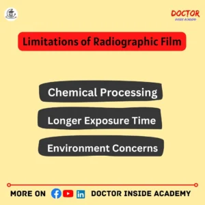
- Chemical Processing
The x ray films were created by developing and fixing films, so these x ray films needed chemical processing is a time-consuming process.
- Longer Exposure Time
The exposure is a little bit longer than digital radiography and is challenging because patients need to be stull at the time of exposure for better and clearer images with motion or blurry.
- Environment Concerns
The chemical used for film processing required proper disposal and handling management, to reduce the environmental risk hazards.
Knowing the limitations of radiographic film is very important in making a decision, on which system is suitable for that diagnostic center.
Digital Radiography
After the concerns of traditional radiographic films, digital imaging or digital radiography came into the role and benefited over traditional ones.
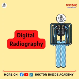
Digital radiography is the advanced version of medical imaging, used to capture different images. These have digital detectors, in place of films inside, and they convert digital data into visuals on the computer.
Digital radiography is more beneficial than traditional one; as it takes less time with easy post-processing and reduces patient radiation dose also.
Digital radiography is more efficient and very quicker in presenting an x ray image with easy post-processing features and reduces radiation exposure for patients.
Digital radiography cannot use X-ray cassettes as it uses indirect flat directors or CCDs (Charged Coupled Devices), known as digital image capture devices, as these directly transfer the image to the system and will convert it into an X-ray image very effectively.
Darkroom procedures are overcome after the existence of digital radiography in medical imaging technology as it bypasses chemical processing and development.
Advantages of Digital Radiography
Digital radiography has benefits over traditional radiographic films, here are a few advantages of digital radiography are followings:
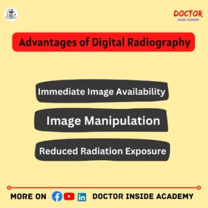
Immediate Image Availability
The main key advantage of digital radiography is to capture the images very quickly, as the exposure is done, and also best to capture real-time images for instant clinical decisions.
Image Manipulation
Digital radiography can be manipulated after image post-acquisition, this flexibility enables radiographers to adjust the image contrast, and image brightness with proper magnification and becomes a very enhanced digital tool in medical imaging.
Reduced Radiation Exposure
Digital radiography takes very little time to capture the x ray images, this saves patients from prolonged exposure and saves them from radiation exposure with reduced risk factors.
Shorter Exposure Time
The exposure time is reduced in digital radiography, and the carrying of x ray cassettes is also reduced, which will be accounted for to save time again.
So, digital radiography is a standard in many diagnostic centers or hospitals, but the radiographic film system is asio continues in specialized medical applications, where digital technology cannot be available.
Types of Digital Radiography
Digital radiography is also further divided into many types based on their use and materials used, are the followings;
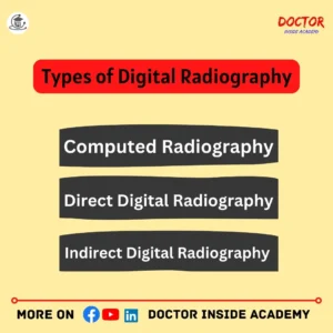
Computed Radiography (CR)
Computed Radiography is used instead of traditional radiography because of its benefits an imaging play]ted is reusable, no darkroom or chemical is accounted for, and software-based reporting and evaluation with simple digital data exchange and archiving.
Direct Digital Radiography (DDR)
Direct Digital radiography(DDR) contains Amorphous Selenium(a-Se); that converts x-ray photons into charge and then a TFT array is used to read these charges and proceed further.
Indirect Digital Radiography (IDR)
Indirect Digital Radiography contains Amporghous Silicon(a-Si).
These use the scintillator layer, made up of cesium iodide(SsI) or gadolinium oxysulfide that converts X-ray to light photons.
Now, this light is directed through a photodiode, converted into electrons, and then further converted into digital signals and seen on the computer.
So, digital radiography uses digital detectors while traditional radiography uses manual x-ray cassettes, which will increase the time and manual laborer, a little bit more.
Last Words
Radiographic films occupy an important space in the world of medical imaging.
This comprehensive chapter of x ray course series serves as a tribute to the essential role of radiographic film in medical imaging and its lasting impact on the field of radiology. While the transition to digital imaging is ongoing, radiographic film remains a symbol of innovation, reliability, and diagnostic excellence in healthcare.
Disclaimer
The above information is based on the author’s learning and experience. If you want to use the above information, then proper citation or mention must be done. Any external link or source may be changed, and we are not responsible for that statistics or data.
I hope you learned something new today.