
X-rays, also known as X-ray radiation, are known for their properties, such as having no mass and no charge.
X-rays are classified based on their energy level, shown by the voltage between electrodes. In this article, we are going to discuss common factors of X-rays, where we discuss radiographic density, quality, artefacts, and so on.
Soft X-rays
Soft X-rays have low-energy photons and longer wavelengths, and the energy of soft X-rays is below 5 keV.
Soft X-rays have very little penetrating power, so they are easily absorbed by low-density tissues such as soft tissues or liquids. Soft X-rays are used to capture soft tissues, like mammography or dental X-rays.
Properties of Soft X-rays
Here are the different properties of soft X-rays are the following:
- The Frequency range of soft X-rays is between 30 and 3000 PHz.
- The wavelength range of soft X-rays is 10 to 0.1 mm
- The level of photon energy is low in soft X-rays.
- Soft X-rays are used in most applications, such as mammography.
So, soft X-rays have a specific range of energy in the X-ray spectrum and are widely used in medical imaging for studying and visualising different structures and abnormalities.
Hard X-rays
Hard X-rays have high-energy photons, above 5-10 keV, and can easily penetrate high-density tissues like bones or metals and are not easily absorbed by lower-density tissues.
Hard X-rays are used in X-rays and computed tomography.
Along with this, hard X-rays are used for industrial purposes, for testing purposes of different metals or objects, and also used in security and at airports for scanning cargo and baggage inspection.
Properties of Hard X-rays
Hard X-rays also have their unique properties, like soft x rays following:
- The Frequency range of hard X-rays is between 3 and 300 EHz.
- The wavelength of hard X-rays is between 100 and 1 pm.
- The photon energy level is higher in hard X-rays, i.e., above 5 to 10 keV.
- The main application of hard X-rays is to image inside objects, such as medical radiography and airport security, and to determine crystal structures, etc.
So, hard X-rays have high-energy photons and are used in medical imaging and also for different purposes.
Difference between Soft and Hard X-rays
Soft and hard X-rays have their unique characteristics and functions; a few of them are here:
The main difference between soft and hard X-rays is that they have different ranges of frequencies, as harder x rays are greater frequencies than soft X-rays.
The second difference is that hard X-rays have higher energy, and Soft X-rays have lower energy.
The third difference is that hard X-rays have shorter wavelengths, whereas soft X-rays have longer wavelengths.
Here, these three differences are enough to differentiate between soft and hard X-rays.
Absorption of X-rays
The absorption of X-rays is the interaction of X-rays with different matter, through the processes known as photoelectric absorption and Compton scattering.
X-ray penetration is one of the X-ray properties, and X-rays can pass through most body tissues or densities and penetrate the body based on their frequency and wavelength. Here, X-ray absorption contributes to X-ray image formation.
We see different shades on the X-ray images due to the different levels of X-ray absorption, and the level of X-ray absorption depends upon the atomic structure, energy level of rays, and density of the materials.
Here, the binding energy of the material is much less than the X-ray energy, so the game of absorption takes place.
High-energy X-ray photons can penetrate high-density substances, such as bones or metals, and vice versa; the level of X-ray absorption directly contributes to the image contrast, and here different levels of absorption properties appear as dark and bright areas on the X-ray image.
Iodinated contrast agents are also used to enhance X-ray absorption and image visibility and diagnosis.
Why is X-ray Absorption Important?
When we diagnose X-ray images for different abnormalities, we see that different body parts appear in different shades of grey. These shades make the image bright or dark, based on the level of X-ray absorption. Dense tissues can’t allow X-rays to pass easily, and vice versa, contributing to X-ray image contrast.
Hence, X-ray absorption shows different levels of X-ray contrast and is used in image diagnosis, helping in identifying pathologies. And also used in scientific research and much more.
Radiographic Density
The radiographic density is the level of darkness or lightness of the X-ray image, which we also call a “radiograph”.
The radiographic density is also called radiodensity.
When the exposure is taken, the amount of x ray photos emitted also influences the density of the X-ray image, which may be overexposed or underexposed.
The level of X-ray attenuation directly depends on the density of the targeted body area; the higher the density, the more X-ray attenuation, or vice versa.
The X-ray image has different density ranges shown as back, white, and grey.
Five X-Ray Densities
When X-ray images are taken, five X-ray densities play a role in the role of making the X-ray image easily readable.
Low-density body areas are seen in black on the X-ray image, and high-density body areas are seen in white on the X-ray film, because of the different densities of the body parts captured in the X-ray.
Here are the 5 most common X-ray densities, are following:
From these five X-ray densities, four X-ray densities are natural, and the rest is metal.
Air
Air is seen on the X-ray image because air is a radiolucent substance. Such as the lungs.
Fat
Fats are seen as lighter than air, such as fat tissue.
Soft Tissue/Water
Fluid fills cavities in the body, seen as whiter than the fat tissues, as they have higher tissue density in that area.
Bone
Human bones are very dense, and very little X-ray can pass through them, so they are seen as white on the X-ray image. Such as calcification of bones, any blockage in the arteries, etc.
Metal
Metal is seen as pure white on the X-ray image as it is a radiopaque material.
The metal is not naturally present in the body, as we already mentioned; it may be implanted in the body, swallowed, or accidentally ingested, such as foreign objects or iodinated contrast media.
Abnormal X-Ray Densities
If you’re a radiologist or radiographer, general physician, you must know these 5 X-ray densities so that you can easily differentiate between normal X-ray densities and abnormal ones, which helps you to diagnose diseases easily and smoothly assist the patient in further evaluation.
Radiographic Quality
Radiographic quality is a very important factor in medical imaging diagnosis, as it shows the clarity of the X-ray image for better diagnostic purposes.
Radiographic quality has many factors, such as image clarity, contrast, resolution, low noise, and desired exposure factors.
If any X-ray image receptor takes more radiation in any specific area, then that area is overexposed and becomes darker in the final X-ray image output, or vice versa,i.e., underexposed.
X-Ray Image Contrast
Contrast is a concept of the difference between radiographic densities, and X-ray image contrast helps radiologists to diagnose abnormalities, such as differentiating between anatomical radiology and pathological radiology.
X-ray image contrast is the result of different densities of the body tissues on the X-ray image. Here, when this density difference is high, the image is known as a high-contrast image.
X-Ray Unsharpness
X-ray unsharpness is a concept of loss of the spatial resolution of the X-ray image and affects the image clarity.
Types of Unsharpness
There are many types of X-ray unsharpness, but here are the main three:
Motion Unsharpness
Motion unsharpness is caused by the movement of the patient.
When an X-ray is taken and the patient moves a little, this will contribute to motion unsharpness.
These patients’ movements may be voluntary or involuntary, depending on the action of the body parts being exposed.
To reduce motion unsharpness, we must keep the patient immobilised or ask the patient not to move and stay still for a whole second or hold their breath for a short time. If we focus on these, then we can easily conquer the motion unsharpness.
Geometric Unsharpness
Geometric unsharpness is the concept of the geometry of the X-ray beam in radiography.
The smaller the focal spot in X-ray, the less geometric unsharpness, and radiographers get the best quality images. This can be captured by OFD (Object-to-Film Distance).
If it is not possible to keep the patient’s body near the X-ray source, then FFD goes beyond the normal range limit and accounts for image unsharpness.
System Unsharpness
The system’s unsharpness is caused by the detector used in the X-ray system because every detector has its own features and characteristics, so they are also limited to some extent.
X-Ray Image Quality
The X-ray image quality results fromX-ray beam penetration power.
mAs and KVP are the two main factors that contribute to the X-ray image; here, mAs results in the number of electrons or photons, and KVP results in the speed of the X-ray beam. So, Proper exposure is a metric that is followed to get better diagnostic images.
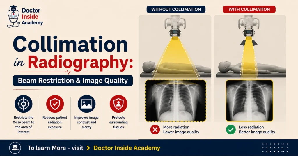
X-ray image quality is influenced by contrast, spatial resolution, detector performance, focal spot size, noise, collimation, artefacts, etc.
While getting better X-ray images, radiographers must be aware of the ALARA Principle. Proper collimation reduces scattered radiation and enhances image quality.
X-Ray Noise
X-ray noise is the random variation of the X-ray photons on an X-ray image and looks like a salt-and-pepper distribution.
That is because of the level of distribution of darker or lighter pixels. This may be high or low. X-ray noise directly contributes to decreasing X-ray image quality and results in poor diagnoses of any diseases or abnormalities.
X-ray images are affected by quantum noise, patients, low radiation dose, motion, and detector quality.
X-Ray Artefacts
X ray artifacts are unwanted shadows in X-ray images and are not accounted for in diagnosis, and degrade X-ray image quality.
The artefacts are outside of the patient’s body. X-ray artefacts may be a result of hardware failure, radiographer errors, software issues, or image processing errors.
The most common causes of X-ray image artefacts are improper X-ray image handling, processing system errors, or patient movements. The most common X-ray artefacts are motion artefacts, detector errors, image processor errors, or grid artefacts.
Last Words
In this article, we learned common factors of X-rays like radiographic density, X-ray image quality, X-ray absorption, X-ray artefacts, and so on.