
In Chapter 6, we will learn collimation in radiography and x ray grids and types of x ray grid in radiology. The x ray course series is for radiology students, where we cover all topics like your friend teaches you.
X Ray Collimator
X-ray collimators are also called beam collimators or beam limiters because they are designed to direct the x-ray beam on the desired area embedded in x-ray tube housing.
This means these limit the field of view of the opening mirror.

The Collimator in the x-ray machine is very important and has lights and mirrors designed to align light and x ray fields.
These two shutters are made up of lead, and with the help of these, we can target radiation in the desired area only.
X-ray collimators resist the scattering and leakage of X-rays and protect the patients from unwanted radiation exposure.
Function of Collimator in X Ray Tube
The function of collimator of the ray beam is to check the x-ray beam and the image quality with better diagnostic results. These are fundamental aspects for better image quality and patient dose.

Here are the main functions of x ray collimators:
Controlled X Ray Beam
X ray beam is limited when proper collimation is entertained during the exposure.
X Ray Collimator prevents the patient from leakage and scattering of x ray beam. These help the radiographers to direct and adjust the x-ray beam.
Improved Image Quality
When the desired x ray beam is targeted on the interest area, as these are collimated, then this gives better image quality by proper absorption of x-rays by tissues and helps radiologists or radiographers to diagnose the abnormalities quickly.
Optimized Result
Proper angulation and x ray beam size are considered when collimation is used, and the result is accurate and easily diagnosed. Here, the larger x-ray beam is needed for bone x-rays, while mammography exams can be taken in a shorter x ray beam.
These are the common function of collimator, as we discussed above.
Types of Collimation
Collimation in Radiography is a very important component of x-ray tube to limit the ray radiation range and decrease radiation exposure for the patients.

There are two types of collimation, which are the following:
Manual Collimation
Radiographers operate manual collimation before the exposure to limit x ray beam by lead leaf with a lever in the x ray tube.
This collimation is done manually every time before the exposure for better diagnosis.
Manual collimation is also classified into many types – NK102, NK202, etc.
Electric Collimation
These are also called automatic collimation and are primarily used in fluoroscopy.
Electric collimation is easily controlled remotely, and there is no need to adjust every time. This is the benefit above the manual collimation.
So, collimation in radiography is very important for better diagnostic images and results.
Types of Collimator in Radiography
Many x ray Collimators in radiography are used according to the requirements and their structures with their principles.
Here are a few main types of collimation techniques, are following:
Fixed Collimators
Fixed collimators are not adjustable and are already set to the range of the desired aperture, where the ray beam comes out easily.
Adjustable Collimators
Adjustable collimators can be adjusted manually, and radiographers can change the x-ray beam direction and area of the x-ray beam to adjust the shape and size of the Collimator’s opening to control the x ray beam.

Parallel Collimators
Parallel collimators have holes and plates designed in parallel to target the beam equally in the desired area.
Focusing Collimators
Focusing collimators are like focusing cups, as these are used to direct the x-ray beam to get the smaller beam spot. These are mostly used in focused directions and high-resolution examinations.
X Ray Grids
When the exposure is taken, radiation becomes scattered and only contributes to the patient radiation dose. In this case, we must check this scattered radiation by following preventive measures.
We use collimators or other devices, including X Ray Grid, to check the scattered radiation. X ray grid filters out the scattered radiation that may be reflected or cause noisy or blurry images and is a component of x-ray machine.
What is Grid in Radiology?
X-ray grids are placed in between the patient and the x-ray cassette.
These are also called filtering devices, used to get better and optimum X-ray images,
X-ray grids are designed in a window format, with an opening, and are a common form of the grate with a narrow strip series of nickel, aluminum, or lead.
Dr. Gustav Bucky invented these x-ray grids in 1913 and explained that these are like a honeycomb.
When the exposure is done, x-rays target the desired area, and these x-rays move randomly. When x-ray grids are used, only straight x rays pass through, and other random x rays are easily absorbed in the way and improve the quality of the x ray image.
Uses of X Ray Grid in Radiology
X ray grids are very important in case of maintaining radiation scattering.
Grids are the most commonly used ix-rays, with different ratio ranges, used as needed, such as 6:1, 12:1, 8:1,10, etc.
Grids are used in different imaging diagnoses, such as the abdomen, spine, skull, IVP, RGU, MCU, mammography, or barium series, to get better images.
Grid Ratio
Grid Ratio shows the working efficiency of the grid and is the ratio of the height of lead strips and the distance between two strips.
Three Dimensions of the Grid
Thickness of lead strips,(T)
Height of strips,(h)
Distance between lead strips,(D)
Grid ratio = h/D

A high grid ratio is better in cleaning radiation scattering, and a high grid ratio is used in high kvP techniques or vice-versa.
The most commonly used high grid ratio is 12:1, and the low grid ratio is 8:1.
A ratio Grid absorbs more scattered radiation than a lower ratio grid.
Advantages of Grid in Radiology
As we learn about X Ray Grid above, here are a few advantages:
Reduces Radiation Scattering
The main purpose of the x-ray grid is to reduce the scattered radiation that only accounts for degrading image quality. These scattered make the image blurry or foggy, which will be misdiagnosed or underdiagnosed. When we use x ray grid, it absorbs these scattered radiations, and the outcome is a better and more detailed image.

Enhances Contrast
When we reduce x ray scattering by using x ray grid, only the desired x ray beam is passed from these x ray grids, which results in improved contrast. This contrast differentiates between tissue densities and opacities.
X ray images with enhanced contrasts help radiologists/radiographers/medical practitioners to diagnose easily and efficiently.
Better Diagnostic Images
The x ray grids result in better diagnostic images, as the scattered radiation is already absorbed. In the case of chest or abdomen examination, x-ray grids are very important as these account for better diagnosis and in better reporting and analyzing the abnormalities and followed further.
Focused X Ray Beam
X ray grids are aligned parallelly and produce radiation scattering. Here, the concept is that x ray beam is passed through strips, as these are designed in a way that goes directly and creates a focused beam, resulting in better image quality.
Disadvantages of Grid in Radiology
Every coin has two sides – a head and a tail; likewise, advantages and disadvantages are two factors for everything. Here are the disadvantages of x ray grids:
Increases Patient Dose
X ray grids absorb x rays themselves, and exposure is increased at the appropriate level for better imaging when they pass through it. The dose increment is limited and follows safety considerations.

Grid Artifact
When x-ray grids are not aligned on x-ray 0image receptors, this creates artifacts on the x-ray image as lines or patterns and causes difficulty in image diagnosis. Proper grid alignment is important in this case; otherwise, it results in grid artifacts.
Costly
As we know, anything that adds extra has additional costs; in this case, x ray grids are additional parts of the x ray machine. When grids are implanted, this cost also accounts for the overall cost. X ray technicians are also trained to use these grids, their alignment, positioning, etc. Nowadays, added in basic radiology courses are added.
Limited in Portable Imaging
X Ray Grids are bulky and not easily used in potable or bedside imaging modalities. So, there are very few portable medical imaging modalities.
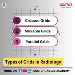
Types of Grids in Radiology
X ray grids are divided into two main parts based on their designs, and the main purpose is to reduce radiation scattering and give better images, but these are different in their use and structure.
1. Stationary Grid
2. Movable Grid
Stationary Grid
Stationary x ray grids are fixed and do not move during exposure. Hence, these are mounted permanently in a grid cabinet and positioned between x ray tube and image receptors.
These grids are used where no movement is allowed.
Types of Stationary Grids
Stationary Grids are also classified based on their structure and use, as follows:
Parallel Grids
Parallel grids are mounted parallel to each other and used for short field sizes or long distances.
Parallel grids only reduce scattering in the grid line directions.
Focused Grids
Focused grids are linear or crossed and comprise lead strips that move slightly toward the focal spot.
These grids are most common and angled to resonate with the divergence of the x ray beam.
Crossed Grids
Crossed grids are two superimposed parallel grids and are very effective in removing radiation scattering.
These grids are also called the Criss-Cross Grid or Cross-Hatch Grid.
Linear Grids
Linear grids are parallel in their longitudinal axis.
Movable Grids
Movable grids are also called Potter Bucky Grid, as named by the inventor Dr. Hollis Potter in 1920.
These oscillate when x ray is taken and run by an electric motor and blurs the shadow of the lead stripes.
Mostly x ray tables have inbuilt linear or focused grids.
The choice between stationary and movable x ray grids is based on the diagnostic center and their specification and examinations taken at that center.
So, radiographers are the main types of grids in radiology and there are many more that we will cover in the upcoming chapter.
Last Words for X Ray Course
We learn about collimation in radiography and x ray grids and different types of grids in radiology. I hope you learn something new today.
Disclaimer
The information provided in this article is for educational purposes only. You can use this content with proper attribution credits to our website.
This article content is based on the learnings and experiences of the author and external links may be changed or updated, so take it as per your efforts.