
In the series, We learn about the history and production of X-rays, their introduction and properties, the principles of radiology, x-ray imaging techniques, imaging evaluation, quality control, quality assurance, patient care and safety in radiography, legal and ethical issues, and future trends in radiography in this series.
So, this is chapter 2 of an x-ray course for paramedical students.
Discovery of X-Rays

- The discovery of x-rays was accidental and changed the world and gives second lives to many people,
- X-rays can see inside our body without any incisions,
- X-rays were discovered by WC Roentgen in 1895,
- He spends most of his time in his lab and working alone, in the 19th century, he was experimenting with Crooke’s tube, (a sealed glass tube with two electrodes, and a vacuum inside, when current passes it shows fluorescent glows),
- One day while working on Crooke’s tube, he noticed that when current passes through the tube, a substance placed a little away from the tube, starts glowing (Barium Platinocyanide),
- This shocked him and then he start experimenting, but the glow was still there,
- This time he covered the tube with black paper, but the glow was still there,
- He did many experiments on this new radiation with many materials there, and found that this radiation passed from these materials, except a few ones only,
- Now, he named them X-Rays, because of their unknown nature at that time,
- Now, he took his wife’s hand x-ray and the images showed the wedding ring and called as 1st Roentgenogram,
- At that time, properties were not discovered, few people were exposed to him for a minute to hours,
- In 1897, ill effects of x rays were seen, on the skin, hair loss, and cancer, and many people died from overexposure,
- WC Roentgen was awarded the first Nobel Prize in 1901 in physics,
- WC Roentgen denied patenting his discovery, if he patents this, this will be limited only to scientific aspects,
- Nowadays, these x-rays are used in medical fields, airports, research and development, engineering, etc.
Components of X-Ray Tube
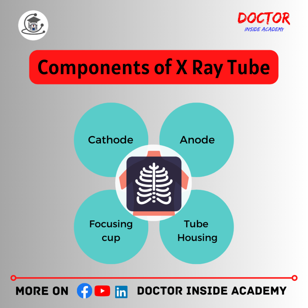
- The primary X-ray source is an X-ray tube,
- The X-Ray tube is an energy converter, here; kinetic energy is converted into X-ray radiation and heat (99% heat and 1% x-rays),
- Among the Components of x-ray tube include cathode, anode, tube envelope, tube housing, cable sockets, x-ray generator, tube window, expansion bellows, filters, collimators, tube envelope, tube housing, x ray port, etc,
- In addition, the exposure unit is powered by a generator, which supplies voltage to the cathode and anode, and the exposure unit.
Cathode
- also known as Filament,
- negative terminal,
- the main source of electrons,
- made up of Tungsten* mainly,
- thermionic emission takes place here,
*We use Tungsten because of its
- high melting point, (3370’C);
- high atomic number (74);
- high thermionic emission property,
- High thermionic emitter,
Cold Cathode
- commonly referred to as a Crooke’s tube or a gas tube that does not need electricity to heat itself,
- an external electric field generates electrons; the generation of x-rays depends on the gas in the room; if the MA changes, a new tube will be fitted; the output is low with unstable production of x-rays, and exposure variables cannot be independently adjusted,
Disadvantages
- include the fact that it consumes a lot of thermal energy and makes the gadget hotter.
Hot Cathode
- also known as the “Coolidge’s Tube,” is a modified cold cathode tube that emits electrons by thermionic emission while also heating the electric filament with a separate current,
- prototype for a contemporary x-ray machine: vacuum inside,
Anode
- – also known as Target,
- – positive terminal,
- – made of copper, molybdenum,
- – where electrons strike,
- – x-ray generated here,
- – it radiates heat and x rays,(99%,1%),
- – Rhenium(10%/5% composition), is used here, to prevent cracking of the anode,
Two types of Anodes
Stationary Anode
- mainly used in dental and portable,
- used when high tube current and voltage are not required,
Rotatory Anode
- used daily machine in the x-ray room,
- it gives a larger target area,
- Made up of tungsten target, molybdenum or graphite mainly,
- High tube current and short time are possible after this,
Motor
- used to rotate the anode,
Rotor
- central iron rod,
- is constructed from copper and mild iron bars,
Stator
- surrounded the rotor, the outside part,
- when current is passed through it, it produces a rotating magnetic field,
Glass Envelope
- Made up of Pyrex,
- sealed and high vacuum,
- Assembles the tube components,
- Provides insulation,
Tube Housing
- it gives shielding and cooling of the x-ray tube,
- housing contains a layer of oil,
- lead shielding is also there,
Collimators
- shape and limit the size of the x-ray beam,
- x-ray field is adjusted,
- it reduces radiation dose to the patients,
Focusing Cup
- focus electrons towards the anode,
- made of molybdenum,
- have a high melting point, with poor thermionic emission,
Other Components are
- expansion Bellows,(space for the oil to expand),
- tube window,( made of Beryllium mainly); low atomic no. and low absorption,
Construction of X Ray Tube
The Construction of X Ray Tube is explained in this detailed video with a well labeled diagram.
To learn more, watch the video below.
Production of X Rays
- The current is passed through the tungsten filament, this filament now heated up due to thermionic emission, and releases electrons, now these electrons are accelerating towards the anode and there they are a collision, and then x rays are produced,
- Fast-moving electrons have kinetic energy and when this energy hitted on target of the anode, this converts into heat and X-rays; (1% X Rays 99% Heat),
- Firstly, current is given to the cathode. It gives electrons due to thermionic emission,
- Now, current is passed through the anode to the cathode,( Electric current moves in the opposite direction of electrons ),
- Here, when current is given to the anode to cathode, due to potential difference, these electrons start moving towards the anode,
- These electrons get sucked on target of the anode, and here, kinetic energy is converted into heat and x rays,
- Shorter wavelength x rays, called Hard X-Rays,
- Longer wavelength x rays, called Soft X-Rays,
Bremsstrahlung Radiation
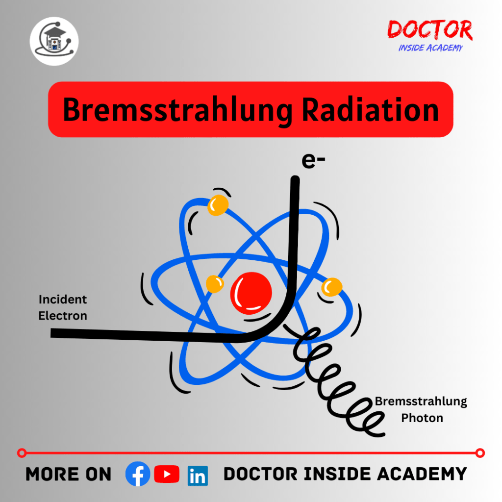
- Additionally known as general radiation or braking radiation,
- and very common in production of x-rays,
- In this; electrons passing near the nucleus get deflected, due to force of attraction,
- As the results; electrons loses its kinetic energy and forms bremsstrahlung radiation,
- The amount of radiation produced, is depends upon the distance between the electron and the nucleus,
- Because they are drawn to the positively charged nuclei in the atoms in the anode, electrons lose kinetic energy as they pass through them.
The closer to the nucleus the electron passes, the more kinetic energy it loses, and it is deflected to continue moving in another direction at lower energy or stopped altogether. The anode emits an X-ray at this point, when the greatest amount of kinetic energy has been transferred.
Characteristic Radiation
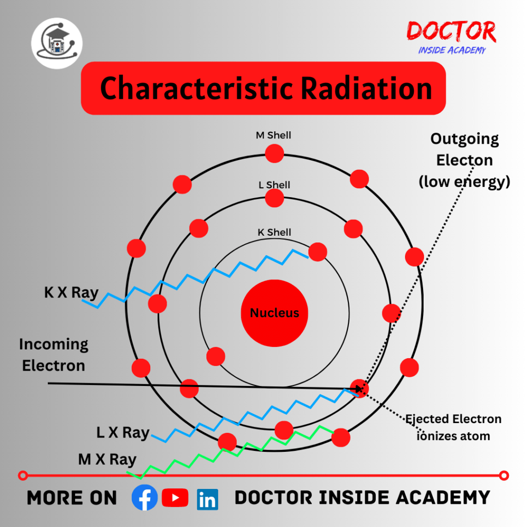
- This produces when the fast-moving electron collides with the k shell,
- Here, after the ejection of electrons, there is a hole behind there,
- Now, the outer shell electrons will fill the void space, where energy is produced, which is known as characteristic radiation,
- If the vacancy is filled with L shells, then α rays,
- If the vacancy is filled with M shells, then β rays,
- If the vacancy is filled with N shells, then α rays,
Electrons are likely to be ejected from the atom if they have an energy equal to or greater than the binding energy of the orbiting electrons in the target atoms.
The inner electron shell, known as the K-shell, is where this primarily occurs.
A photoelectron is the name given to the expelled electron. According to the principle of energy conservation, the K-shell vacancy must be filled for the atom to remain stable, therefore outer shell electrons descend to fill the shell. The name comes from the fact that the x-rays produced by this process of electron transfer between shells are “characteristic” of the binding energies of that specific atom or substance.
Cold and Hot Cathode

- WC Roentgen discovered x rays in 1895,
Cold Cathode
- Commonly referred as crooke’s tube or gas tube,
- Doesn’t need electricity to heat itself,
- External electric current produces electrons and the production of electrons depends on the gas present in the tube,
- If the MA changes, new tube will be fitted;
- Output is low and unstable production of x-rays,
- Exposure variables cannot adjusted independently in this tube,
The main disadvantage is; it consumes a lot of thermal energy and makes the device hotter,
Hot Cathode
- Also known as coolidge’s tube,
- Is a modified version of cold cathode tube,
- Produces electrons by thermionic emissions,
- Heating of filament from separate current is required,
- Vacuum inside,
The prototype for the new modern x-ray machines.
Stationary Anode
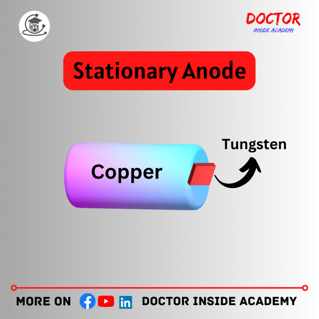
- Target of the tube, where x rays are produced,
Stationary Anode
- It stays stills, called a fixed anode,
- Anode is made up of copper with a small tungsten plate embedded in it; tungsten is the target here,
- Length 1 mm and thickness 2 mm,
- The anode angle is at 15 to 20%,
- The focusing cup prevents the tube wall from electron bombardments,
- The stationary anode x ray tube has a small target area, which limits heat dissipation,
- If the heat keeping strikes on the point repeatedly, it deforms the anode and reduces the efficiency and image quality of the x-rays produced,
- It lowers x-ray output,
- Are used in dental x rays, portable fluoroscopy, or in portable units, etc,
Rotatory Anode
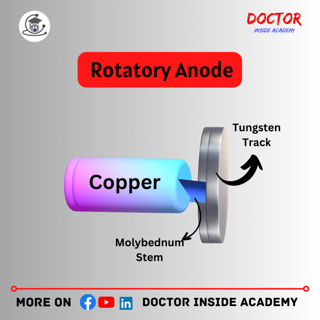
Rotatory Anode
- It is a small metal disk made-up of copper or tungsten that receives an electron from the cathode and produces X-rays,
- It spins around a fixed point,
- Consists of Tungsten and Rhenium (large disc),
- As we know that heat produces in large amount, if heat heats the same point in the surface of an anode can deform and changes the working of the anode,have greater heat loading,
- But in the case of a rotating anode, the heat is distributed equally on the surface of the entire anode. This gives long life to the anode and performs better imaging,
- It takes longer exposures and heavy doses also,
- It has higher output,
Parts of Rotating Anode
- It consists of Rotor, Stator, Anode stem;
Rotor
- Anode connected to the rotor,
- Made up of copper with tungsten disc,
- It rotates by the stators,
Anode Stem
- Made up of Molybdenum and a short one,
Stator
- It is a set of electric coils that produce a strong magnetic field around the glass tube, which helps to rotate the rotor,
- Also known as an induction motor,
Anode Angle
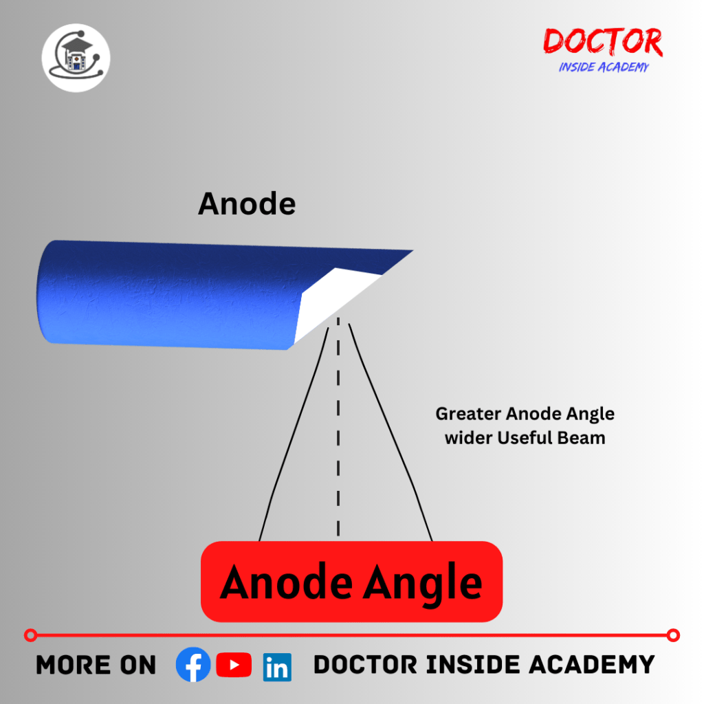
- Is the angle between the anode and cathode,
- Electrons collide from cathode and produces x rays,
- represented by; θ,
- It is about 12 to 15%,
Effectiveness Of Anode Angle
- Anode Angle increases;
- Effective focal spot size increases, or vice versa,(smaller angle results in the effective focal spot).
Final Words
In this chapter 2 of X Ray Course, we learned about discovery of x-rays, production of x-rays and bremsstrahlung and characteristic radiation or difference between stationary and rotating anode x ray tube and much more,
I hope you enjoyed this article and get towards your academic career and you can connect with us on all social media.
Disclaimer
The information provided in this article is only for educational purposes. We keep the information up to date and accurate based on different studies, you can use this article content with proper mentioning, and misuse of the content is strictly prohibited.
The article content is based on learning, experiences, and research performed by the author and we don’t provide specific guidance and recommendations at an individual level.
This article may contain external links to other websites or resources. We have no control over the concrete availability.
Don’t follow any medical advice given on this blog, without consulting with your doctor or physicians first.


Pingback: X Ray Tube Protection and Their Importance - X Ray Course: Chapter 5 - Doctor Inside Academy
Pingback: All About X Ray Tube - X Ray Course: Chapter 3 - Doctor Inside Academy