In this chapter, we talk about x ray tube overload protection, beam centering devices, and beam limiting devices and their types and importance.
Benefits of X-Ray Tube Overload Protection Circuits
X ray tube overload protection circuits play a very important role in the performance of the x ray machine.
There are many overload protection devices available in the market, according to the demand these are used accordingly.

Now, we discuss a few benefits of this are following:
Enhanced Patient Safety
By preventing potential tube damage or failure, overload protection circuits minimize the risk of unexpected malfunctions during X-ray examinations, and help in the protecting patients from unnecessary radiation exposure.
Extended X-ray Tube Lifespan
Properly implemented protection circuits help prolong the life of the X-ray tube by preventing damage caused by thermal stress and electrical overloads.
Consistent Image Quality
By maintaining the X-ray tube within safe operating limits, these protection circuits contribute to consistent and reliable image quality in medical imaging studies.
X-ray tube overload protection circuits are safety features in modern medical imaging systems.
They serve as guardians, continuously monitoring critical parameters and intervening promptly to prevent potential damage to the X-ray tube.
By ensuring patient safety, extending the tube’s lifespan, and preserving image quality, these protection circuits play a vital role in advancing the field of radiology and improving patient care.
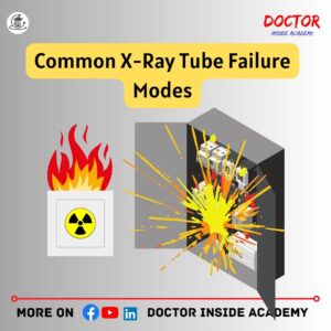
Common X-Ray Tube Failure Modes
Heating of x ray tube is very common when prolonged radiation exposure is taken.
So, preventing x ray tube from this excessive heat is very important, to maintain x ray tube and its performance.

Here are 9 factors of x ray tube failure to keep in mind to increase the lifespan of x ray tubes, are following:
Normal Aging
X ray tubes have limited life because of their material used and characteristics.
The x-ray tube degrades and output decreases until the optimum quantity is not perfect. So, use it effectively and proper exposure slows x ray tube aging.
Filament Burnout
Thermionic emission takes place at the filament side and produces an electron cloud, that is further used in x ray production.
The x-ray tube becomes hotter when more tube current is demanded from the x-ray tube at a fixed voltage, in this case, this filament may burn out.
To avoid filament burnout, radiographers must ensure the optimum temperature in the x ray room.
Slow Leaks
X ray tube needs a high vacuum inside to produce desired x-ray beams, Here, glass to metal seals may allow minute gas to enter, increasing the gaseous pressure.
This leads to the degradation of tube performance.
Inactivity
X ray tubes must be used regularly.
Otherwise, this may allow gases to enter the x ray tube vacuum and cause disability of x ray tube to produce adequate x rays.
Glass Cracking
Most tube contains glass as the wall material.
It insulates x ray tube from leakages.
But, with time, depending on the usage of the x-ray unit, both electrode surfaces start evaporating causing tube leakages and leading to glass breakage in the x-ray tube.
Accidental Damages
Accidental damages are mostly influenced while manufacturing or installing the x ray unit.
Sometimes, misunderstanding, false assumptions, or unfamiliarity with the systems leads to accidental damage.
Temperature
Heat is the enemy of x ray tube so that x ray tube cooling is maintained to keep the x ray tube safer.
If x ray unit is operated at exceeded temperature limits may lead to x ray tube failure or x ray tube cracking.
Dielectric Oil Leakages
This oil prevents x ray tube from cracking.
This oil consumes heat produced in the x-ray tube and is put out from the system to keep that safe.
As this oil is heated again and again, may lead to burnout of x ray tube glass and leads to x ray tube burnout.
Overheating
This may cause tube burnout or may explode the x ray tube housing.
Mostly x ray units have a fan and pump system to prevent from x ray tube overheating.
Sometimes, dust also inhibits the natural air and becomes a cause of x-ray tube overheating.
Hence, these are 9 x ray tube failure factors. Make sure to check all these factors to keep x ray tube long-lasting and well-maintained.
Beam Centering Devices
Beam Centering Devices are the devices used for proper patient positioning because these show the direction of central ray light from the x-ray beam from the x-ray tube to the cassette area.
These are also called collimating devices or collimators.
These are present in x-ray tube with mirror and lights made of lead shutters and helps to focus the radiation according to the area of interest.

If the x ray beam is not centered or aligned, then it captures extra body areas, then suggested and will be completely irrelevant for diagnostic purposes and leads to misaligned and misdiagnosis.
Importance of Beam Centering Devices
Beam-centering devices are very important and play great in imaging. Because these are designed to ensure the focus of the x-ray beam on the area of interest.
Here, are a few importance of beam centering devices, are followings:
Minimizing Radiation Exposure to Non Targeted Areas
Beam centering devices have a adjustable aperture that checks the x ray beam, by narrowing or widening the aperture, radiographers control the size of the x-ray beam and target it to the area of examination.
Better Beam Alignment
Beam centering devices are designed with the best alignment mechanisms that help radiographers to rotate the x ray tube in multiple directions. These movements help in better centering the x-ray beam on the desired surfaces.
Improving Patient Safety
Beam centering devices directly help in radiation protection as these stop unwanted radiation exposure. Proper lead shields and collimations ensure better diagnostic images with less scattering of radiation and leakage.
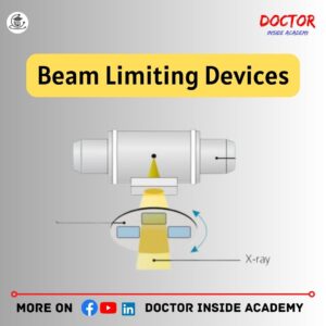
Beam Limiting Devices
Beam limiting devices also called collimators, is a device that is designed to narrow down the primary x ray beam and saves patients from unwanted radiation exposure.

Beam limiting devices are directly connected to the x-ray tube and refine the x ray beam before hitting the body for x ray images.
Importance of Beam Limiting Devices
Beam limiting devices are designed to shape the x ray beam on the area of interest, also known as the field of view (FOV).

Along with this here are a few importance beam limiting devices, are following:
Reduces Patient Radiation Exposure
Beam limiting devices limit the x-ray beam, so, radiation is limited to the area of interest and prevents the patient from unwanted radiation exposure.
Better Image Quality
When beams narrow down, only limited radiation is directed to the desired area, and image clarity and sharpness are enhanced.
Reduces Radiation Scattering
Radiation scattering lower the image quality and increases the radiation exposure to the nearby body tissues.
When these x ray beams are limited and shaped by these collimators or beam limiting devices, this saves body tissues from unwanted radiation.
Follows ALARA Principle
ALARA – “As Low As Reasonably Achievable”, this is a basic fundamental principle of radiation safety. All these beam limiting devices strictly follow the ALARA principle, to maintain radiation safety and patient safety.
So, these are the main importance of beam limiting devices.
Types of Beam Limiting Devices
Beam limiting devices are a very crucial component of radiography systems. These can control unwanted x ray radiation and shape the x ray beam.

There are many types of beam limiting devices, a few of them are the following:
Aperture Diaphragms
The aperture diaphragm is made up of a lead sheet with a center hole, that is used to determine the size and shape of the x ray beam.
Aperture diaphragm connected to x ray tube directly.
The main disadvantages of aperture diaphragms are fixed shape and size and reduced sharpness and resolution.
Cones
Cones are metal tubes, directly connected to x ray tube, and used to limit the shape and size of the beam.
Cones are also the modification of apertures and are made up of lead.
Extension Cylinders
Extension cylinders are about 10 to 20-inch metal tubes in a cylindrical shape and mostly connect to the cones to increase the length of the cone.
The main advantages of cones and cylinders are better beam restriction than aperture diaphragms.
These both also reduce scatter radiation.
Collimator’s
Collimators are very common type of x ray beam limiting device.
Collimators are made up of two lead shutters inside the x ray tube housing.
These shutters are independently adjustable in the shape of squares and rectangles.
Collimators have an inbuilt light, that is very useful for radiographers to adjust the collimation and direct the x ray beam to the area of interest accurately.
Grids
X-ray grid is a very important part of the ray system. These filters unwanted radiation from the x ray beam.
X-ray grid is a device that is used to enhance the images.
X-ray grid is made up of lead, nickel, or aluminum narrow strips, that stop the x-rays.

These are also called radiographic grid.
The main purpose of grids is to reduce scatter radiation and enhance the quality and image contrast to give better images by removing scattering of radiation.
Along with Grid, mAs, and KvP are also the major factors of image quality.
Radiographic Film
Radiographic films give x-ray images for diagnostic and screening purposes. These are made from many layers, one of them is the emulsion layer, which is responsible for latent image.
The film processing develops latent images into visible film.

These are the x-ray film and gives us the diagnostic result for the exposures taken in the radiography. These are the printouts of the patient’s diagnoses.
There are different types of x-ray films used in the day-to-day radiography outputs.
Automatic Exposure Control (AEC)
Automatic exposure controls are the devices used in x ray receptors, for terminating the exposure, when the appropriate amount of radiation is detected in these systems.
Automatic exposure control is used in radiographic and mammographic imaging modalities.
Simply, in radiography, these devices are placed before the x-ray image receptors.

These contain photomultiplier tubes, ionization chambers, or solid-state detectors.
The main advantages of Automatic exposure control are to give better consistent densities, reduce dose repeats.
Final Words
In this Chapter 5 of the X Ray Course, we learned about beam limiting devices and beam centering devices with their importance and their types with advantages and disadvantages.
You can connect with us on all social media, and I hope you enjoyed this article as you move forward with your academic career.
Disclaimer
The information in this article is solely for educational purposes. We keep the material up to date and correct based on numerous studies. The content of this article may be used with due attribution, but its misuse is prohibited.
The article content is based on the author’s knowledge, experiences, and research, and we do not offer specific advice or suggestions at an individual level.
This page may contain external connections to other websites or resources. We have no control over the concrete supply.
Take no medical advice from this website without first consulting your doctor or physicians.


