
The skin is one of the organs, cavities, arteries, and tracts that are lined by tissue. Tissue epithelial cells are resistant to abrasion and possess a special ability to heal damage. Discover every aspect of epithelial tissue, including its kinds, functions, structure, and more.
There are four types of animal tissues: connective, muscular, nerve, and epithelial tissues. Since epithelial tissue covers both the interior and exterior body surfaces, it is the most widely distributed tissue.
Understanding Epithelial Tissue
The epithelium, also known as epithelial tissue, lines the body cavity and forms the skin’s outer layer. It makes up the lining of the digestive, reproductive, respiratory, and excretory systems. They carry out many tasks, including secretion, sensing, absorption, and defense.
The tissues of the epithelium are almost entirely avascular. For example, nutrients must reach the tissue by diffusion or absorption from the surface or underlying tissues since blood vessels cannot pass through the basement membrane.
Numerous epithelial tissues have the ability to quickly replace dead and damaged cells. Surface epithelium’s ability to shed injured or dead cells enables our digestive tracts and airways to quickly replace damaged cells with new ones.
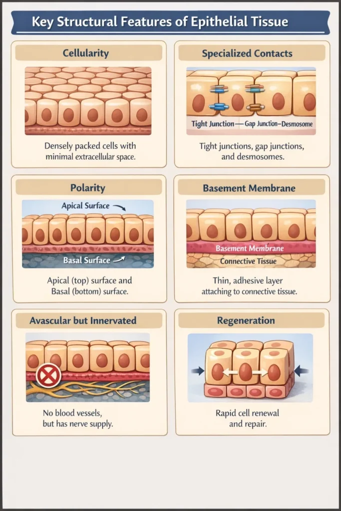
Key Structural Features of Epithelial Tissue
A continuous, closely packed layer of cells forms epithelial tissue. The epithelium tissue has one surface that is exposed to bodily fluids or the outside world. A membrane made of fibers and polysaccharides released by epithelial cells connects the other surface to the tissue.
A few particular “design rules” enable epithelial tissues to function as the body’s main barrier.
Here are the key structural features:
- Cellularity: Nearly all of them are composed of closely spaced cells with very little matrix (intercellular material) between them.
- Specialized Contacts: Tight junctions (leak-proofing), gap junctions (communication channels), and desmosomes (rivets for strength) are examples of specialized junctions that keep cells together.
- Polarity: There is a “top” and a “bottom.” The basal surface is connected to the underlying tissue, whereas the apical surface confronts the exterior or a bodily cavity.
- Basement Membrane: All epithelial sheets are attached to the connective tissue underneath them by a thin, non-living adhesive layer known as the basement membrane.
- Avascular but innervated: They lack avascular blood vessels. They absorb nutrients from deeper tissues by diffusion. But since they are innervated, that is, fed by nerve fibers, you can experience touch and pain.
- Regeneration: They have a high capacity for regeneration and may quickly replace damaged cells through cell division since they are frequently subjected to friction or environmental harm.
Classification by Number of Layers
Based on the number of cell layers piled on top of one another, epithelial tissue is divided into three primary groups. The behavior of the tissue is directly determined by its structure.
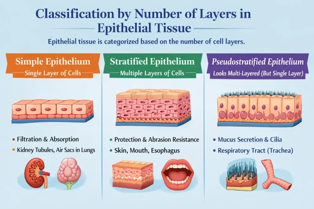
Single Layer Simple Epithelium
- The basement membrane is connected to a single layer of cells. It is ideal for swiftly moving molecules, but its thinness makes it poor for protection.
- Filtration, secretion, and absorption are the main functions.
- It can be found in the kidney tubules for blood filtration, the intestines for nutritional absorption, and the lining of the air sacs (lungs) for gas exchange.
Stratified Epithelium (Multiple Layers)
- This is made up of two or more cell layers. The basement membrane is only touched by the deepest layer. Its primary function is that of a “bodyguard.”
- Main Purpose: Defense against chemical or physical deterioration (abrasion).
- Where to find it: The outer layer of your skin (epidermis), the lining of your mouth, and the esophagus. As the top layers get rubbed off, new cells from the bottom push up to replace them.
Pseudostratified Epithelium (The “Fake” Layer)
- The word “pseudo” denotes falsity. Because the cell nuclei are at varying heights, this seems to have several layers, although each and every cell really contacts the basement membrane. In theory, there is only one layer.
- Mucus secretion and movement (frequently with microscopic hairs called cilia) is the primary function.
- The majority of the upper respiratory system (trachea), which aids in capturing and expelling dust and debris from your lungs, is where you may locate it.
Classification by Cell Shape
The physical characteristics of epithelial cells are used to classify them. Consider them as various kinds of “building blocks” intended for particular purposes.
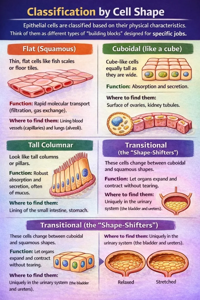
Flat (squamous)
- These are thin, flat cells that look like fish scales or floor tiles. They enable quick dispersion since they are so thin.
- Function: Quick molecular transport (filtration, gas exchange).
- Where to locate them: lining the blood arteries (capillaries) and lungs (alveoli).
Cuboidal (like a cube)
- They resemble dice or cubes and are about the same height as they are wide. Their additional “bulk” gives organelles space to create or process materials.
- Function: Absorption and secretion.
- They can be seen on the surface of the ovaries and in the kidney tubules.
Tall Columnar
- These appear to be towering columns or pillars. Their height offers additional defense and room for intricate cellular components (such as Golgi structures and mitochondria).
- Function: Strong absorption and secretion, usually of mucus.
- They are located in the lining of the small intestine and stomach.
Transitional (the “Shape-Shifters”)
- This is a special kind of cell that adapts its form to the demands of the body. The cells appear cuboidal when the tissue is relaxed and squamous when it is stretched.
- Function: To allow organs to expand and contract without tearing.
- Where to locate them: Exclusively in the urinary system (the bladder and ureters).
Specialized Types of Epithelial Tissues
Although the majority of epithelial tissues fit into the conventional “shape and layer” classifications, two specialized varieties are made for extremely particular mechanical and chemical tasks.
The “Shape-Shifters” or Transitional Epithelium
- This tissue is a master of disguise. The cells in this stratified (multi-layered) epithelium alter their appearance depending on the degree of stretch.
- Appearance: The top cells seem plump and cuboidal when the organ is empty. They become squamous and flatten down when the organ is full.
- Function: It prevents organs from rupturing or leaking when they expand and contract.
The “Secretors” of the glandular epithelium
- These are epithelial cells that have been “reprogrammed” to secrete particular compounds, such as sweat, hormones, or enzymes. They are categorized into two main groups:
The “Producers” or germinal epithelium
- From a purely biological perspective, the reproductive system has a specific layer of cuboidal cells.
Examples of Epithelial Tissues
To make this easy to visualize, here are the most common examples of epithelial tissues categorized by their specific “job” in the body:
- Simple Squamous: Found in the air sacs of the lungs (alveoli) and the lining of the heart and blood vessels (endothelium). Its thinness allows oxygen to pass through instantly.
- Simple Cuboidal: Forms the walls of kidney tubules and the surface of the ovaries. These cells act like little pumps for secretion and absorption.
- Simple Columnar: Lines the stomach and intestines. These tall cells often have “microvilli” (tiny folds) to grab as many nutrients as possible from your food.
- Stratified Squamous: Your skin (epidermis) is the classic example. It’s also found in the mouth, esophagus, and vagina. These areas take a lot of friction, so they need “disposable” top layers.
- Stratified Cuboidal/Columnar: These are rare. You’ll find them in the large ducts of sweat glands, mammary glands, and salivary glands.
- Pseudostratified Columnar: Lines most of the upper respiratory tract (trachea). These cells usually have cilia (tiny hairs) that act like a broom to sweep mucus and dust out of your airways.
- Transitional: Found in the urinary bladder and ureters. These cells are unique because they can “morph” from plump to flat as your bladder fills up.
- Exocrine: Sweat glands, salivary glands, and pancreas (releasing digestive enzymes through ducts).
Endocrine: Thyroid gland and adrenal glands (releasing hormones directly into the blood).